Chromosomal disorders
Chromosomal disorders: You can study chromosomes using karyotyping. In karyotyping, chromosomes are stained and visualized under a microscope during metaphase. After Giemsa staining, chromosomes show characteristic banding patterns. GC-rich regions stain dark with G banding, bright with R banding, and dark with Q banding. Karyotyping can detect abnormalities in chromosome number (e.g., trisomy 21) and chromosome structure (e.g., inversions). However, microdeletions and mutations are too small to be seen on karyotyping; these can be detected using FISH (fluorescence in situ hybridization).
-
Numerical abnormalities of chromosomes: An abnormal number of chromosomes (aneuploidy) can occur in two main ways:
- Monosomy: an individual is missing one chromosome from a pair.
- Trisomy: an individual has more than two copies of a chromosome instead of a pair.
Triploidy is the presence of three sets of chromosomes (a total of 69 chromosomes) and results in miscarriage. The most common cause of aneuploidy is nondisjunction during meiosis I.
Disorders caused by aneuploidy involving autosomes and sex chromosomes
| Aneuploidy | Disorder |
| Trisomy 21 (Down syndrome) | Most cases from nondisjunction during meiosis, others from robertsonian translocation between chromosomes 14 and 21 or mosaicism; higher risk if maternal age > 35 years; mental retardation, prominent epicanthal folds, single palmar crease or simian crease, higher risk of Alzheimer’s disease and ALL, CHD and GI abnormalities |
| Trisomy 18 (Edwards syndrome) | Mental retardation, VSD, prominent occiput, receding jaw, low set ears, club foot, clenched fists |
| Trisomy 13 (Patau syndrome) | Mental retardation, cleft lip and palate, extra digits or toes, clenched fists, club foot, holoprosencephaly, CHD, urogenital defects |
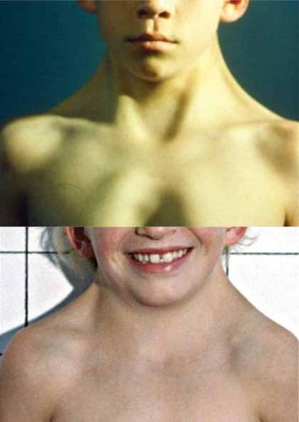
| 47 XXY, 48 XXXY (Klinefelter syndrome) | Tall stature, disproportionately long arms and legs, hypogonadism, undescended testes, gynecomastia, infertility, hypospadias, psychosocial problems, higher risk of breast cancer and SLE, genotypically and phenotypically male |
| 47XYY syndrome | Tall males, learning disabilities, hypotonia, may show aggressive behavior |
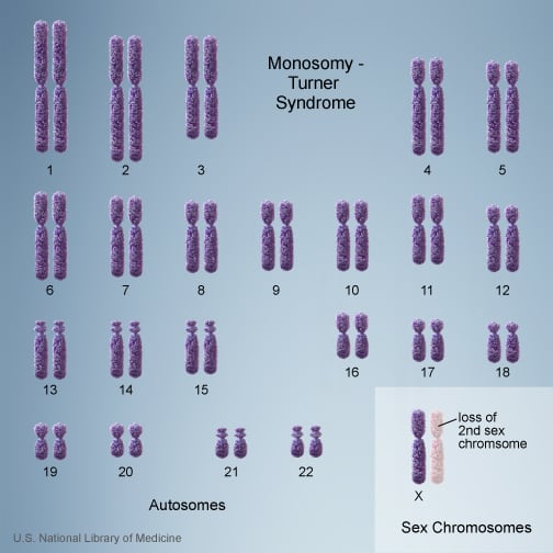
| 45 XO (Turner syndrome) | Short stature, webbed neck, widely spaced nipples, streak ovaries, premature ovarian failure, lymphedema of the hands and feet, coarctation of aorta, skeletal abnormalities; monosomy X or mosaicism |
-
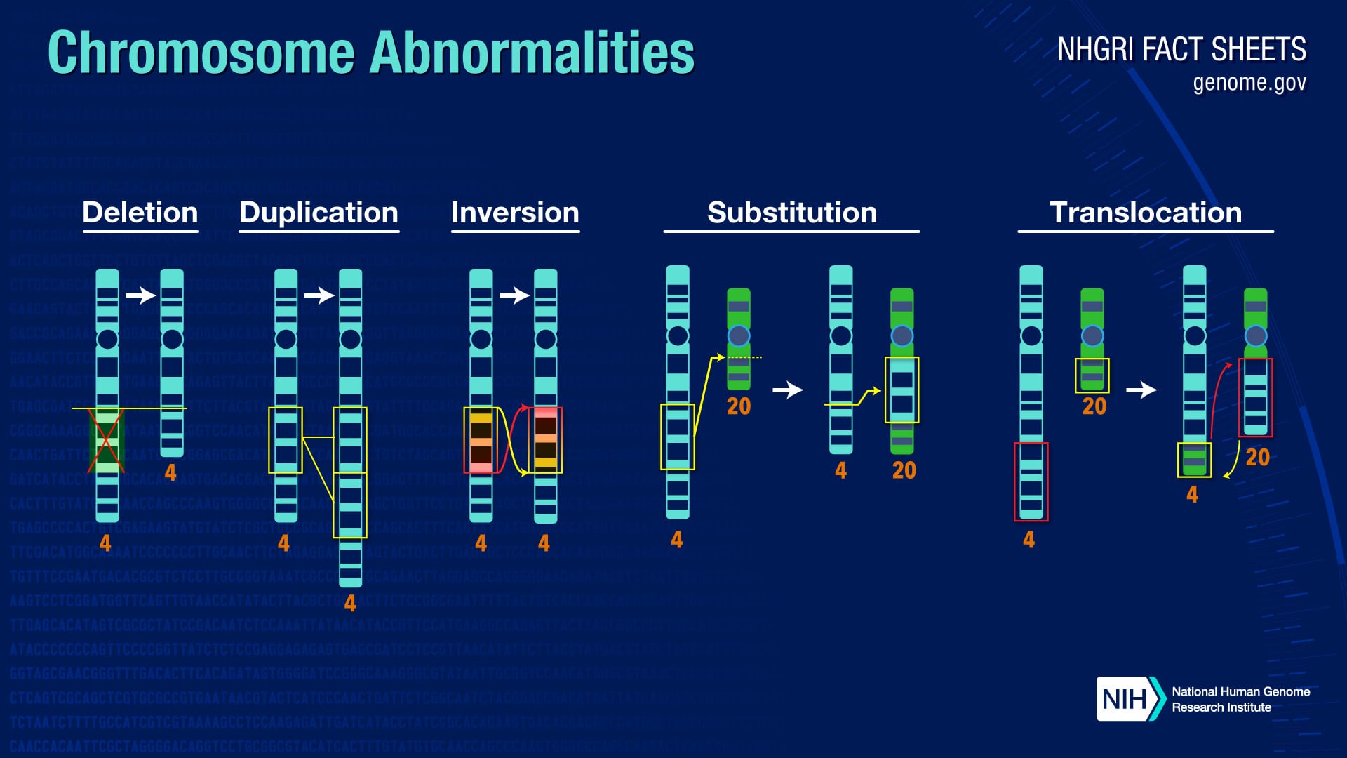
Structural abnormalities of chromosomes: These result from events that cause a loss or gain of genetic material, including the following.
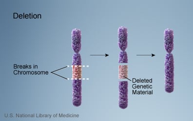
Deletions: A portion of the chromosome is deleted (lost). This is seen in Cri du chat syndrome, in which there is deletion of part of the 5p chromosome. It is characterized by cat-like cry, mental retardation, microcephaly, epicanthal folds, low set ears, small jaw and widely spaced eyes. Many cancers show a deletion of the Tp53 tumor suppressor gene-containing region of chromosome 17p.
Microdeletions (deletions where <3 megabases are lost) may occur due to unequal crossing over between homologous chromosomes with multiple repeat DNA sequences. Disease-producing microdeletions are seen in Prader Willi and Angelman syndromes, neurofibromatosis type 1, Charcot Marie Tooth disease, DiGeorge syndrome, etc.
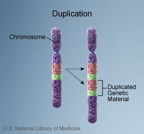
Duplications: A small part of a chromosome may be duplicated along its length, increasing the number of genes present.
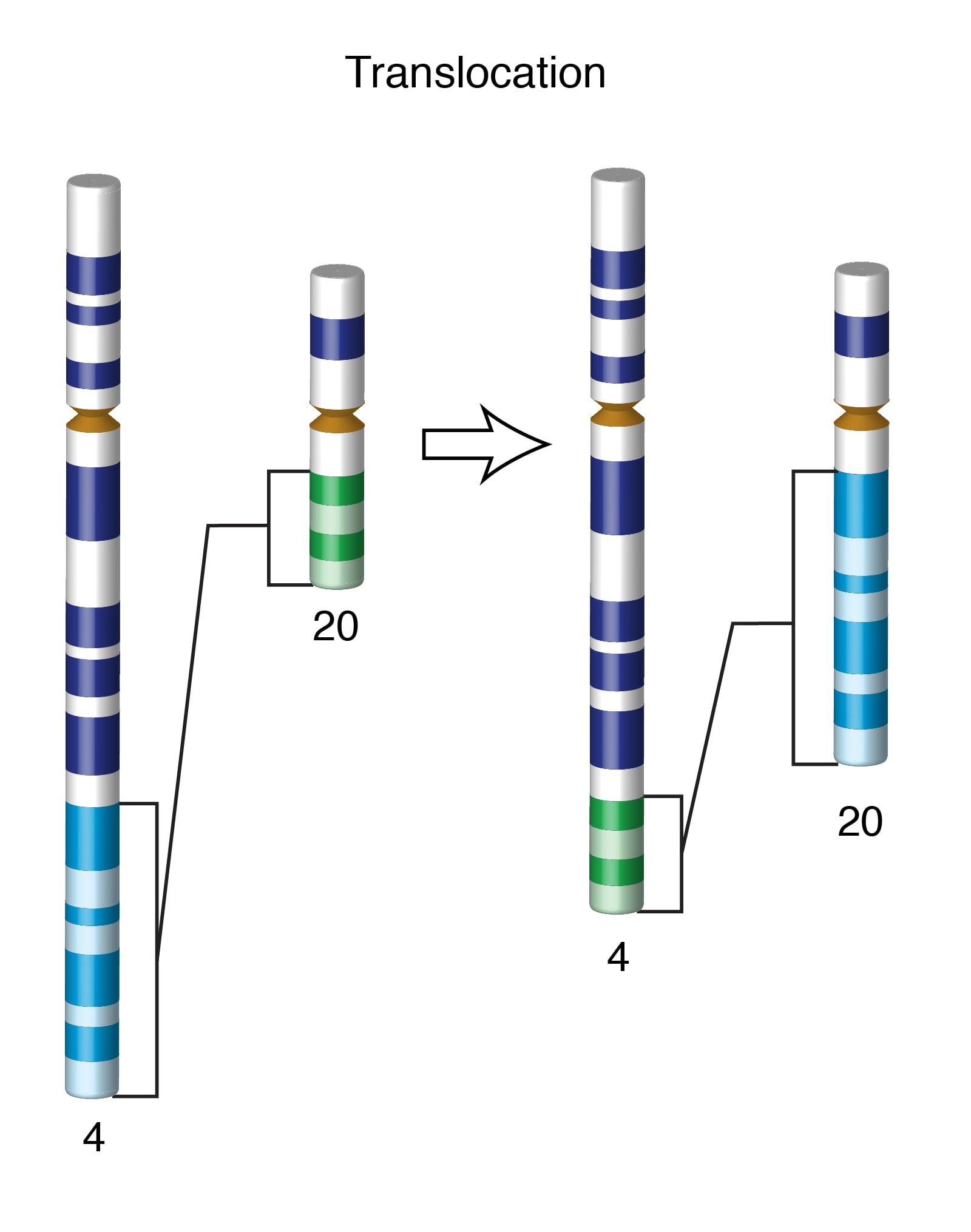
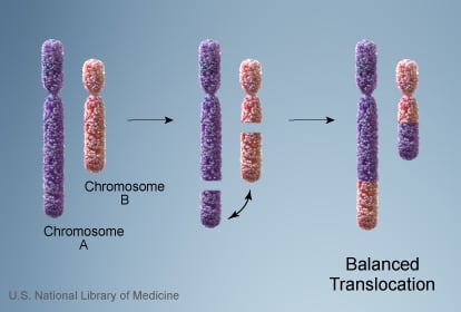
Translocations: This is a rearrangement of chromosome material involving two or more chromosomes. Translocations may be reciprocal or Robertsonian.
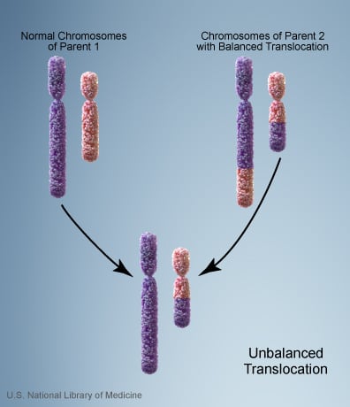
Reciprocal translocation occurs when genetic material is exchanged between two non-homologous chromosomes. It is often balanced, meaning there is no net gain or loss of genetic material after the translocation. Carriers of balanced translocations may be normal but have an increased risk of miscarriages or having offspring with congenital defects. Translocations in somatic cells may give rise to cancers; for example, the Philadelphia chromosome t(9;22) causes CML.
Robertsonian translocations occur during translocation of acrocentric chromosomes, when there is a loss of the short arms and fusion of the long arms. During meiosis, Robertsonian translocations may cause trisomies and monosomies.
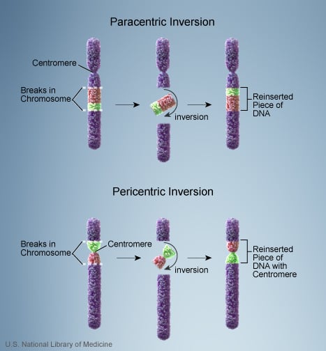
Inversions and ring chromosome:
- Inversion: a portion of a chromosome breaks off, turns upside down, and is then reattached to the chromosome.
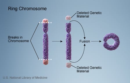
- Ring chromosome: forms when chromosome ends attach to form a ring. Offspring may receive an imbalance of chromosomal material.
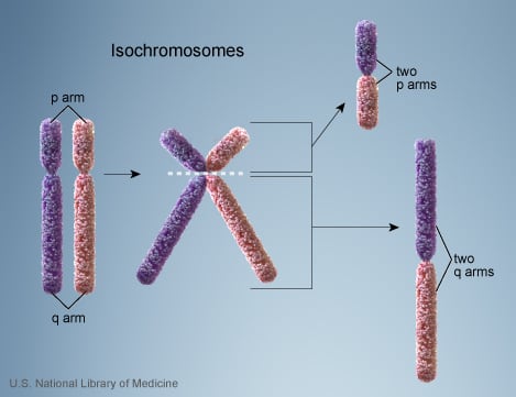
Isochromosome: This is an abnormal metacentric chromosome formed by duplication of one arm of a normal chromosome with deletion of the other arm. Both arms of the new chromosome look like mirror images of each other.