



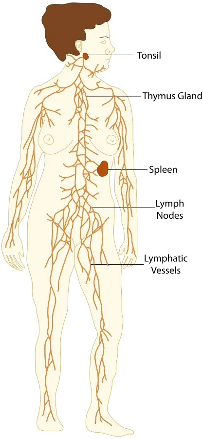
Anatomy of the immune and lymphatic systems
When working with allergies and infectious diseases it is important to be familiar with the lymphatic and immune systems. They are two different systems; however, they cooperate and work together to maintain homeostasis. They also defend the body from foreign pathogens, which can cause infectious diseases. The cells, tissues, and organs of the lymphatic and immune systems are almost the same. But the two systems help the body in very different ways. The lymphatic system can be described as mostly structural. It is the physical component of the two systems. The immune system can be described as mostly functional. The cells of the immune system work to keep the body safe from pathogens. These two systems together make up an amazingly complex and efficient means of maintaining the body’s health.
The lymphatic system is responsible for a number of functions with the help of the immune system. Lymphatic functions include cleansing the cellular environment, returning proteins and tissue fluids to the blood, providing a pathway for the absorption of fats into the bloodstream, and defending the body against disease.
Immunology is the healthcare specialty that deals with many immune- and lymphatic-related diseases and disorders. An immunologist is a specialist involved in the diagnosis, treatment, and prevention of disorders of the immune and lymphatic systems. Other healthcare specialists who work with these diseases and disorders include cardiologists, allergists, and rheumatologists.
Anatomy of the immune and lymphatic systems
The immune and lymphatic systems are composed of many structures that allow these two systems to function and protect the body. The immune system involves white blood cells to help protect against pathogens. The following sections discuss the anatomy and physiology of the immune and lymphatic systems.
Anatomy of the immune system
Two very important cells in the immune system, monocytes and lymphocytes, pass from the bloodstream through the blood capillary walls into the spaces between the cells in body tissue. When they pass into the lymph that surrounds cells, they perform their protective functions. Monocytes and lymphocytes are covered in the white blood cell sections that follow; lymphocytes are much more complicated and, because they are essential to the immune response, are discussed in a later section of this chapter.
Cells of the immune system
The immune system is composed of organs, tissues, cells, and chemical messengers. The components of the immune system interact to protect the body from external invaders and the body’s own internally altered cells. The chemical messengers are cytokines. They are secreted by cells of the immune system that direct immune cellular interactions. Most of the cells involved in the lymphatic and immune systems are white blood cells (WBCs). A quick review of white blood cells may help in the discussion of the immune system.
Granular white blood cells
The three types of granular WBCs are neutrophils, eosinophils, and basophils. They are characterized by their heavily granulated cytoplasm and segmented nuclei. Neutrophils are aggressively phagocytic - that is, they engulf and destroy invading pathogens. Eosinophils are involved in allergies. Basophils play a part in inflammation.
Unlike red blood cells (RBCs), WBCs are found in both the bloodstream and the tissues. During inflammation, the blood carries neutrophils through blood vessels to the site of injury. Capillary walls become more permeable, and the granular cells squeeze through to the site of infection. Once at the site of infection or injury, the neutrophils engulf the invading microorganism. The collection of WBCs, dead WBCs, bacteria, and tissue cells creates pus at the site of infection.
Agranular white blood cells
The two types of agranular leukocytes are monocytes and lymphocytes. They have a clear cytoplasm (no granules) and a solid nucleus. Monocytes are large and become macrophages when they enter tissues. Macrophages are also aggressively phagocytic cells that engulf pathogens and debris. Lymphocytes are small cells that have a big job! They are responsible for much of the work in specific immunity. Lymphocytes are further classified into T lymphocytes and B lymphocytes, depending on their function in the immune response. B lymphocytes and T lymphocytes do not look different. But they function differently. B and T lymphocytes are also called B cells and T cells.
Anatomy of lymph system
Interstitial fluid is the fluid that bathes the cells in body tissue. Once this fluid enters the lymphatic vessels, it is called lymph. Lymph flows through the lymphatic system, which is composed of these structures:
- Lymph vessels, lymph nodes, lymph glands, and lymphoid tissue
- Lymph organs: the tonsils, adenoids, vermiform appendix, spleen, thymus gland, and Peyer patches
Lymph moves in one direction. This prevents pathogens from flowing through the entire body. The system filters out the microorganisms as the lymph passes through various capillaries, vessels, and nodes.
Physiology of the immune and lymphatic systems
The best way to understand the immune system is to learn about the body’s various levels of defense. The goal of foreign pathogens is to enter the body, reproduce, and damage healthy tissue. The immune system’s primary function is to differentiate what is “self” from what is “foreign” and then destroy anything that is foreign. The immune system wants to stop pathogens from causing harm. The body’s defenses can be pictured as three layers: two layers of nonspecific immunity (also known as innate immunity), followed by a layer of specific immunity (also known as adaptive immunity), which can be natural or acquired. Most pathogens can be contained by the first two lines of nonspecific defense. However, some pathogens get past nonspecific defenses. These pathogens are met with the third line of defense, or specific immunity.
Nonspecific immunity
The term nonspecific immunity refers to the many ways the body protects itself from pathogens without having to “recognize” them. The first line of defense in nonspecific immunity consists of the following methods of protection.
- Physical: Examples include intact skin and mucous membranes. These are physical barriers to pathogens. The skin protects the inside of the body from the outside environment. The sticky mucous membranes at many body openings trap pathogens.
- Reflexes: Examples include coughing, sneezing, vomiting, and diarrhea. These actions help get rid of pathogens by forcing them back out of the body.
- Chemical: Examples include tears, saliva, and perspiration. These have a slightly acidic pH, which discourages pathogens from entering the body. All these fluids also help wash the pathogens away. In addition, stomach acids and enzymes try to kill pathogens.
The second line of defense in nonspecific immunity goes to work if the pathogens make it past the first line of defense. The second line of defense uses cellular and chemical responses to destroy the pathogen. The following are the protective measures of the second line of defense:
- Phagocytosis: The process of cells engulfing and destroying invaders. Pathogens that make it past the first line of defense and enter the bloodstream may be consumed by neutrophils, monocytes, or macrophages.
- Inflammation: A protective response to irritation or injury. Signs and symptoms of inflammation include heat, swelling, redness, and pain. This process causes a brief vasoconstriction, followed by vasodilation and an increase in vascular permeability. The resulting increase in blood flow and fluid leakage into the tissue produces the heat, redness, and swelling of inflammation, creating a good environment for healing. If caused by a pathogen, the inflammation is called an infection.
- Pyrexia: The medical term for fever. When an infection is present, fever helps protect the infected area. Fever increases the action of phagocytes and reduces the viability of certain pathogens. Most pathogens love normal body temperature, so when a fever is present, the environment gets too hot, and they cannot function as they should.
- Protective proteins are part of the second line of defense. These include interferons, which disrupt viral replication and limit a virus’s ability to damage cells. Complement is a group of proteins made in the liver that plays a key role in the immune response, tagging pathogens for destruction (opsonization) and directly damaging their cell membranes. Complement proteins are inactive in the blood until they encounter bacteria. Meeting bacteria activates complement, enabling it to lyse (destroy) the organisms.
- Natural killer (NK) cells are the last cell type involved in the second line of defense. They are derived from stem cells in the bone marrow, concentrated in the liver and lungs, and involved in nonspecific immunity and inflammation. NK cells are also capable of destroying targeted cells, such as tumor cells and virus-infected cells. They are not phagocytic cells. This special kind of lymphocyte acts nonspecifically to kill cells that have been infected by certain viruses and cancer cells.
Specific immunity
Specific immunity is different from nonspecific immunity in several ways:
- Specific immunity means that the immune response is generated against one specific antigen. The response will not be effective against any other antigens.
- Specific immunity counts on the immune cells to identify antigens and recognize them if they encounter them again. An antigen can be an infectious agent from outside the body (exogenous), or it can be a malignant or tumor cell inside the body (endogenous). Both types of antigens are foreign, something that is not normal to the immune system.
- Specific immunity prepares a specific response (antibody production) to that unique antigen.
- Specific immunity intensifies with repeated challenges by the same antigen. When the body encounters a known antigen, it can respond more efficiently and react to the antigen quickly.
- Specific immunity may be either genetic or acquired. Genetic immunity is an inherited ability to resist certain diseases because of one’s species or individual genetic makeup - it is present from birth rather than learned through antigen recognition and memory. Acquired immunity, in contrast, develops as the body encounters and responds to specific antigens.
The two types of antigens and the two methods of specific immunity are summarized below.
Antibodies belong to a group of proteins called immunoglobulins (Igs). There are five distinct classes of immunoglobulins: IgG, IgM, IgE, IgA, and IgD. Each class of immunoglobulin responds to different types of immune challenges, or antigens.