


Achievable logoAchievable logo
CCMA
Sign in
Sign up
Purchase
Textbook
Practice exams
Support
How it works
Exam catalog
Mountain with a flag at the peak
Textbook
1. Medical assistant
2. Electronic records
3. Medical terminology and anatomy
4. The fundamentals of infection control
5. Introduction to vital signs
6. The patient interview and history
7. The physical examination
8. Appointment scheduling
9. Insurance billing
10. Diagnostic coding and the ICD-10-CM System
11. Procedural coding
12. Medical billing and reimbursement essentials
13. Assisting with medical specialties
14. Assisting with the musculoskeletal system
15. Assisting with the cardiovascular system
16. Assisting with the respiratory system
17. Assisting with the nervous system
18. Anatomy and physiology of the urinary system
18.1 Urinary system anatomy
18.2 Urinary system diseases and disorders
18.3 Chronic kidney disease and nephrotic syndrome
18.4 Male reproductive system anatomy and physiology
18.5 Medical assistant's role in urinary examinations and treatments
19. Assisting in obstetrics and gynecology
20. Assisting in endocrinology
21. Assisting in ophthalmology & otolaryngology
22. Assisting in gastroenterology
23. Assisting in the immune & lymphatic systems
24. Assisting in pediatrics: the developmental stages and care
25. The medical assistant’s role in caring for the older patient
26. The role of the medical assistant in physical therapy examination and assessment
27. Preparing for minor surgery: room, solutions, and supplies
28. Introduction to the clinical laboratory
29. Urinalysis
30. Blood collection
31. Analysis of blood
32. Electrocardiography and heart structure
33. The principles of pharmacology
34. Essential calculations and measurement systems
35. Solid, liquid, & solutions medication doses
36. Administering medications
37. Metabolism and core nutrient roles
38. Medical emergencies in the healthcare setting
Achievable logoAchievable logo
18.1 Urinary system anatomy
Achievable CCMA
18. Anatomy and physiology of the urinary system
Our CCMA course is currently in development and is a work-in-progress.

Urinary system anatomy

8 min read
Font
Discuss
Share
Feedback

Urology is the healthcare specialty that deals with most urinary diseases and disorders. A urologist is a specialist involved in the diagnosis, treatment, and prevention of disorders of the urinary system and the male reproductive system. Nephrology is the healthcare subspecialty that focuses on kidney function and disorders. A nephrologist diagnoses and treats kidney conditions.

Besides the urology department, patients with urinary and male reproductive concerns are typically seen in primary care, urgent care, and even specialty departments. Medical assistants should have a strong understanding of the common urinary signs, symptoms, diseases, diagnostic tests, and treatments. Sexually transmitted infections (STIs), which can impact both the urinary and male reproductive systems, are discussed in another chapter.

The anatomy, physiology, and pathology of the urinary system will be discussed before the male reproductive system is addressed.

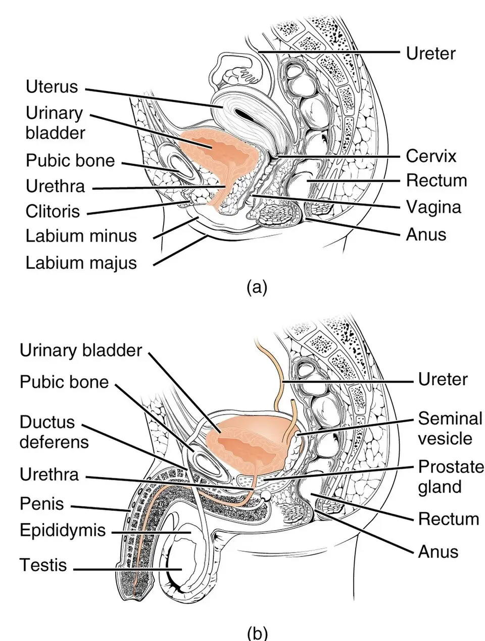
The urinary system is composed of two kidneys, two ureters, a urinary bladder, and a urethra. The kidneys filter the blood and eliminate waste through the passage of urine. The ureters move urine from the kidneys to the bladder. The urinary bladder stores the urine until it is excreted. The urethra is the tube that conducts the urine out of the bladder.

Female and male urinary systems compared
Female and male urinary system
Wikimedia Commons
/
CC BY 3.0

Kidneys

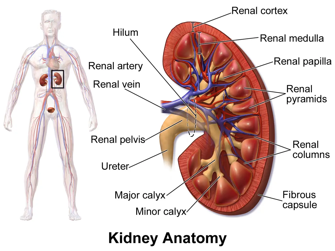
The kidneys are bean-shaped organs that are the size of a fist. The hilum (an indentation) is the location on the kidney where the ureter and renal vein leave the kidney and the renal artery enters. The kidneys are located posterior to the peritoneum , the muscles of the back, and between the T12 and L3 vertebrae. The left kidney is situated about 1 inch (2 cm) higher than the right because of the location of the liver.

The kidneys remove unwanted substances from the blood and form urine for excretion. Blood is delivered to the two kidneys by the renal arteries, which branch off the abdominal aorta. Blood flows from the renal arteries into afferent arterioles (smaller arteries) and through a network of capillaries called the glomeruli. Filtration of the blood occurs in the glomerulus. Blood from the glomerulus moves into the efferent arterioles and into the peritubular capillaries. As the blood leaves the kidneys, it moves from the peritubular capillaries to venules and then empties into the renal veins. The renal veins take the blood out of the kidneys. Blood from the renal veins flows into the inferior vena cava as it heads to the heart to become oxygenated again.

Internal and external parts of the kidney labeled
Parts of the kidney
Wikimedia Commons
/
CC BY 3.0

The kidney structures

If a kidney were sliced open, you would see the following:

  • Capsule: The fibrous outer covering of the kidney
  • Cortex: The outer portion of the kidney
  • Renal column: An extension of the cortex that dips down between the medullary pyramids
  • Medulla: The inner portion that extends from the end of the cortex to the calyces
  • Medullary pyramid: A cone-shaped structure located in the medulla and containing straight tubular structures and blood vessels
  • Minor and major calyces and the renal pelvis: Extensions of the ureter inside the kidney

Nephrons

Each kidney contains tissue with millions of microscopic units called nephrons. Nephrons are the functional units of the kidneys. They are located in the cortex and extend into the medulla. Each nephron is a very long tube or tubule. One end of the nephron is the renal corpuscle, and the remaining section of the nephron is the renal tubule. The renal corpuscle consists of the following:

Blood flow to and from the kidney:

  • Bowman capsule: A cup-shaped structure of the nephron that surrounds the glomerulus
  • Glomerulus: A cluster of capillaries inside the Bowman capsule

The afferent arteriole brings blood into the glomerulus, and the efferent arteriole takes blood away from the glomerulus. The diameter of the afferent arteriole is much larger than the diameter of the efferent arteriole. The importance of the narrowed diameter will be discussed in the Physiology of the Urinary System section of this chapter.

The renal tubule is a long, thin, twisted tube that brings the filtrate (which will become urine) from the Bowman capsule to the renal pelvis. During the passage of filtrate from the Bowman capsule to the renal pelvis, water and electrolytes move between the urine and the blood to maintain homeostasis. The formation of urine will be discussed later in the chapter. The renal tubule is made up of the following:

  • Proximal (convoluted) tubule (PCT): The first section of the renal tubule that is attached to the Bowman capsule.
  • Henle loop: Follows the PCT and includes a straight descending section, a loop, and then a straight ascending section.
  • Distal (convoluted) tubule (DCT): Follows the ascending section of the Henle loop.
  • Collecting duct (CD): Multiple distal tubules join and form this straight collecting duct, which empties into the calyx.

To summarize, the nephron begins on one end with the Bowman capsule, which is filled with capillaries. The proximal tubule is the next section of the nephron. It is followed by the Henle loop, the distal tubule, and finally, the collecting duct. Many collecting ducts empty into one calyx. Many calyces merge into the renal pelvis, which is an extension of the ureter inside the kidney. The Bowman capsule, proximal tubules, and distal tubes are in the cortex. The Henle loop and the collecting ducts are in the medulla of the kidney.

Ureters

The bilateral ureters are thin, muscular tubes approximately 10 inches long. The tube’s muscular layer creates peristaltic waves that help move the urine to the bladder. The ureters’ walls are made of transitional epithelium , which allows the ureters to stretch to accommodate urine flow. Many nerve endings are also in the walls. A stone in the ureter can cause severe pain.

The point where the ureter enters the bladder is called the ureterovesical junction. The ureters enter the bladder at an angle. This creates a flap over the end of the ureter, which serves as a valve. Urine can empty into the bladder, but it cannot back up from the bladder and reenter the ureter. This mechanism also prevents bacteria from moving from the bladder up to the kidneys.

Bladder

The urinary bladder is a hollow organ in the pelvic cavity. It serves as a holding tank for urine. Urine enters the bladder through the ureters and leaves the bladder through the urethra. The triangular area in the bladder between the ureters’ entrance and the urethral outlet is called the trigone.

The transitional epithelium creates the inner lining of the bladder. When the bladder is empty, it flattens, and the walls overlap in folds, also known as rugae. This creates a wrinkled appearance. As the bladder starts to fill with urine, the rugae and transitional epithelium allow for greater bladder volume. Think of a balloon that was blown up and then emptied of air. The balloon is smaller and wrinkly in appearance. When inflated, the wrinkles are gone, and it can accommodate a large volume of air. The same thing happens with the bladder and urine.

The bladder is also composed of smooth muscle fibers that make up the detrusor muscle. The detrusor muscle relaxes when the bladder fills. It contracts to push urine out of the bladder and into the urethra. Normally, the bladder holds about 1.5 to 2 cups (360 to 480 mL) of urine during the day and doubles that amount at night. As the bladder fills to about 150 mL, stretch receptors in the detrusor send a message to the central nervous system. As more urine enters the bladder, the urge to urinate increases. After urination or micturition, residual urine remains (usually less than 50 mL) in the bladder.

Urethra

The urethra is a mucous membrane–lined tube that drains urine out of the bladder. In males, the internal urethral sphincter is located at the bladder end (or proximal end) of the urethra. The sphincter is made of smooth, involuntary muscles. The external sphincter is located in different places in males and females. The external sphincter is made of skeletal muscles and is a voluntary muscle. A voluntary muscle provides you with the ability to control when you urinate. The distal end of the urethra is called the urinary meatus. The urinary meatus is the final structure before the urine leaves the body.

Overview of Urinary System Anatomy

  • Composed of kidneys, ureters, bladder, urethra
  • Kidneys filter blood, form urine; ureters transport urine to bladder
  • Bladder stores urine; urethra conducts urine out of body

Kidneys

  • Bean-shaped, located between T12–L3 vertebrae (left higher than right)
  • Receive blood via renal arteries; filter blood in glomeruli
  • Blood exits via renal veins to inferior vena cava

Kidney Structures

  • Capsule: fibrous outer covering
  • Cortex: outer region; Medulla: inner region with medullary pyramids
  • Calyces and renal pelvis: collect urine, connect to ureter

Nephrons

  • Functional units, millions per kidney, in cortex and medulla
  • Renal corpuscle: Bowman capsule (surrounds glomerulus)
  • Renal tubule: PCT → Henle loop → DCT → collecting duct
    • Collecting ducts drain into calyces, then renal pelvis

Ureters

  • Muscular tubes (~10 inches), move urine via peristalsis
  • Lined with transitional epithelium for stretch
  • Ureterovesical junction prevents backflow into ureters

Bladder

  • Hollow pelvic organ, stores urine (1.5–2 cups daytime)
  • Lined with transitional epithelium and rugae for expansion
  • Detrusor muscle contracts for urination; trigone is triangular area between ureters and urethra

Urethra

  • Mucous membrane–lined tube draining urine from bladder
  • Internal sphincter: smooth, involuntary muscle
  • External sphincter: skeletal, voluntary muscle (controls urination)
  • Urinary meatus: external opening

Physiology of the Urinary System

  • Maintains fluid volume and electrolyte balance
  • Regulates blood pressure (renin secretion)
  • Controls red blood cell production (erythropoietin)
  • Activates vitamin D for calcium/phosphorus absorption

Urine Formation

  • Three steps: filtration, reabsorption, secretion

Filtration

  • Occurs in glomerulus/Bowman capsule (renal corpuscle)
  • High pressure forces water and small solutes (electrolytes, urea, glucose) into Bowman capsule
  • Blood cells and proteins remain in capillaries
  • Low blood pressure reduces filtration and urine output

Sign up for free to take 14 quiz questions on this topic

Previous
Next  | 18.2 Urinary system diseases and disorders
All rights reserved ©2016 - 2026 Achievable, Inc.

Urinary system anatomy

Urology is the healthcare specialty that deals with most urinary diseases and disorders. A urologist is a specialist involved in the diagnosis, treatment, and prevention of disorders of the urinary system and the male reproductive system. Nephrology is the healthcare subspecialty that focuses on kidney function and disorders. A nephrologist diagnoses and treats kidney conditions.

Besides the urology department, patients with urinary and male reproductive concerns are typically seen in primary care, urgent care, and even specialty departments. Medical assistants should have a strong understanding of the common urinary signs, symptoms, diseases, diagnostic tests, and treatments. Sexually transmitted infections (STIs), which can impact both the urinary and male reproductive systems, are discussed in another chapter.

The anatomy, physiology, and pathology of the urinary system will be discussed before the male reproductive system is addressed.

The urinary system is composed of two kidneys, two ureters, a urinary bladder, and a urethra. The kidneys filter the blood and eliminate waste through the passage of urine. The ureters move urine from the kidneys to the bladder. The urinary bladder stores the urine until it is excreted. The urethra is the tube that conducts the urine out of the bladder.

Kidneys

The kidneys are bean-shaped organs that are the size of a fist. The hilum (an indentation) is the location on the kidney where the ureter and renal vein leave the kidney and the renal artery enters. The kidneys are located posterior to the peritoneum , the muscles of the back, and between the T12 and L3 vertebrae. The left kidney is situated about 1 inch (2 cm) higher than the right because of the location of the liver.

The kidneys remove unwanted substances from the blood and form urine for excretion. Blood is delivered to the two kidneys by the renal arteries, which branch off the abdominal aorta. Blood flows from the renal arteries into afferent arterioles (smaller arteries) and through a network of capillaries called the glomeruli. Filtration of the blood occurs in the glomerulus. Blood from the glomerulus moves into the efferent arterioles and into the peritubular capillaries. As the blood leaves the kidneys, it moves from the peritubular capillaries to venules and then empties into the renal veins. The renal veins take the blood out of the kidneys. Blood from the renal veins flows into the inferior vena cava as it heads to the heart to become oxygenated again.

The kidney structures

If a kidney were sliced open, you would see the following:

  • Capsule: The fibrous outer covering of the kidney
  • Cortex: The outer portion of the kidney
  • Renal column: An extension of the cortex that dips down between the medullary pyramids
  • Medulla: The inner portion that extends from the end of the cortex to the calyces
  • Medullary pyramid: A cone-shaped structure located in the medulla and containing straight tubular structures and blood vessels
  • Minor and major calyces and the renal pelvis: Extensions of the ureter inside the kidney

Nephrons

Each kidney contains tissue with millions of microscopic units called nephrons. Nephrons are the functional units of the kidneys. They are located in the cortex and extend into the medulla. Each nephron is a very long tube or tubule. One end of the nephron is the renal corpuscle, and the remaining section of the nephron is the renal tubule. The renal corpuscle consists of the following:

Blood flow to and from the kidney:

  • Bowman capsule: A cup-shaped structure of the nephron that surrounds the glomerulus
  • Glomerulus: A cluster of capillaries inside the Bowman capsule

The afferent arteriole brings blood into the glomerulus, and the efferent arteriole takes blood away from the glomerulus. The diameter of the afferent arteriole is much larger than the diameter of the efferent arteriole. The importance of the narrowed diameter will be discussed in the Physiology of the Urinary System section of this chapter.

The renal tubule is a long, thin, twisted tube that brings the filtrate (which will become urine) from the Bowman capsule to the renal pelvis. During the passage of filtrate from the Bowman capsule to the renal pelvis, water and electrolytes move between the urine and the blood to maintain homeostasis. The formation of urine will be discussed later in the chapter. The renal tubule is made up of the following:

  • Proximal (convoluted) tubule (PCT): The first section of the renal tubule that is attached to the Bowman capsule.
  • Henle loop: Follows the PCT and includes a straight descending section, a loop, and then a straight ascending section.
  • Distal (convoluted) tubule (DCT): Follows the ascending section of the Henle loop.
  • Collecting duct (CD): Multiple distal tubules join and form this straight collecting duct, which empties into the calyx.

To summarize, the nephron begins on one end with the Bowman capsule, which is filled with capillaries. The proximal tubule is the next section of the nephron. It is followed by the Henle loop, the distal tubule, and finally, the collecting duct. Many collecting ducts empty into one calyx. Many calyces merge into the renal pelvis, which is an extension of the ureter inside the kidney. The Bowman capsule, proximal tubules, and distal tubes are in the cortex. The Henle loop and the collecting ducts are in the medulla of the kidney.

Ureters

The bilateral ureters are thin, muscular tubes approximately 10 inches long. The tube’s muscular layer creates peristaltic waves that help move the urine to the bladder. The ureters’ walls are made of transitional epithelium , which allows the ureters to stretch to accommodate urine flow. Many nerve endings are also in the walls. A stone in the ureter can cause severe pain.

The point where the ureter enters the bladder is called the ureterovesical junction. The ureters enter the bladder at an angle. This creates a flap over the end of the ureter, which serves as a valve. Urine can empty into the bladder, but it cannot back up from the bladder and reenter the ureter. This mechanism also prevents bacteria from moving from the bladder up to the kidneys.

Bladder

The urinary bladder is a hollow organ in the pelvic cavity. It serves as a holding tank for urine. Urine enters the bladder through the ureters and leaves the bladder through the urethra. The triangular area in the bladder between the ureters’ entrance and the urethral outlet is called the trigone.

The transitional epithelium creates the inner lining of the bladder. When the bladder is empty, it flattens, and the walls overlap in folds, also known as rugae. This creates a wrinkled appearance. As the bladder starts to fill with urine, the rugae and transitional epithelium allow for greater bladder volume. Think of a balloon that was blown up and then emptied of air. The balloon is smaller and wrinkly in appearance. When inflated, the wrinkles are gone, and it can accommodate a large volume of air. The same thing happens with the bladder and urine.

The bladder is also composed of smooth muscle fibers that make up the detrusor muscle. The detrusor muscle relaxes when the bladder fills. It contracts to push urine out of the bladder and into the urethra. Normally, the bladder holds about 1.5 to 2 cups (360 to 480 mL) of urine during the day and doubles that amount at night. As the bladder fills to about 150 mL, stretch receptors in the detrusor send a message to the central nervous system. As more urine enters the bladder, the urge to urinate increases. After urination or micturition, residual urine remains (usually less than 50 mL) in the bladder.

Urethra

The urethra is a mucous membrane–lined tube that drains urine out of the bladder. In males, the internal urethral sphincter is located at the bladder end (or proximal end) of the urethra. The sphincter is made of smooth, involuntary muscles. The external sphincter is located in different places in males and females. The external sphincter is made of skeletal muscles and is a voluntary muscle. A voluntary muscle provides you with the ability to control when you urinate. The distal end of the urethra is called the urinary meatus. The urinary meatus is the final structure before the urine leaves the body.

Key points

Overview of Urinary System Anatomy

  • Composed of kidneys, ureters, bladder, urethra
  • Kidneys filter blood, form urine; ureters transport urine to bladder
  • Bladder stores urine; urethra conducts urine out of body

Kidneys

  • Bean-shaped, located between T12–L3 vertebrae (left higher than right)
  • Receive blood via renal arteries; filter blood in glomeruli
  • Blood exits via renal veins to inferior vena cava

Kidney Structures

  • Capsule: fibrous outer covering
  • Cortex: outer region; Medulla: inner region with medullary pyramids
  • Calyces and renal pelvis: collect urine, connect to ureter

Nephrons

  • Functional units, millions per kidney, in cortex and medulla
  • Renal corpuscle: Bowman capsule (surrounds glomerulus)
  • Renal tubule: PCT → Henle loop → DCT → collecting duct
    • Collecting ducts drain into calyces, then renal pelvis

Ureters

  • Muscular tubes (~10 inches), move urine via peristalsis
  • Lined with transitional epithelium for stretch
  • Ureterovesical junction prevents backflow into ureters

Bladder

  • Hollow pelvic organ, stores urine (1.5–2 cups daytime)
  • Lined with transitional epithelium and rugae for expansion
  • Detrusor muscle contracts for urination; trigone is triangular area between ureters and urethra

Urethra

  • Mucous membrane–lined tube draining urine from bladder
  • Internal sphincter: smooth, involuntary muscle
  • External sphincter: skeletal, voluntary muscle (controls urination)
  • Urinary meatus: external opening

Physiology of the Urinary System

  • Maintains fluid volume and electrolyte balance
  • Regulates blood pressure (renin secretion)
  • Controls red blood cell production (erythropoietin)
  • Activates vitamin D for calcium/phosphorus absorption

Urine Formation

  • Three steps: filtration, reabsorption, secretion

Filtration

  • Occurs in glomerulus/Bowman capsule (renal corpuscle)
  • High pressure forces water and small solutes (electrolytes, urea, glucose) into Bowman capsule
  • Blood cells and proteins remain in capillaries
  • Low blood pressure reduces filtration and urine output

More from Anatomy and physiology of the urinary system

  • Urinary system diseases and disorders
  • Chronic kidney disease and nephrotic syndrome
  • Male reproductive system anatomy and physiology
  • Medical assistant's role in urinary examinations and treatments