


Achievable logoAchievable logo
CCMA
Sign in
Sign up
Purchase
Textbook
Practice exams
Support
How it works
Exam catalog
Mountain with a flag at the peak
Textbook
1. Medical assistant
2. Electronic records
3. Medical terminology and anatomy
4. The fundamentals of infection control
5. Introduction to vital signs
6. The patient interview and history
7. The physical examination
8. Appointment scheduling
9. Insurance billing
10. Diagnostic coding and the ICD-10-CM System
11. Procedural coding
12. Medical billing and reimbursement essentials
13. Assisting with medical specialties
14. Assisting with the musculoskeletal system
15. Assisting with the cardiovascular system
16. Assisting with the respiratory system
17. Assisting with the nervous system
18. Anatomy and physiology of the urinary system
18.1 Urinary system anatomy
18.2 Urinary system diseases and disorders
18.3 Chronic kidney disease and nephrotic syndrome
18.4 Male reproductive system anatomy and physiology
18.5 Medical assistant's role in urinary examinations and treatments
19. Assisting in obstetrics and gynecology
20. Assisting in endocrinology
21. Assisting in ophthalmology & otolaryngology
22. Assisting in gastroenterology
23. Assisting in the immune & lymphatic systems
24. Assisting in pediatrics: the developmental stages and care
25. The medical assistant’s role in caring for the older patient
26. The role of the medical assistant in physical therapy examination and assessment
27. Preparing for minor surgery: room, solutions, and supplies
28. Introduction to the clinical laboratory
29. Urinalysis
30. Blood collection
31. Analysis of blood
32. Electrocardiography and heart structure
33. The principles of pharmacology
34. Essential calculations and measurement systems
35. Solid, liquid, & solutions medication doses
36. Administering medications
37. Metabolism and core nutrient roles
38. Medical emergencies in the healthcare setting
Achievable logoAchievable logo
18.4 Male reproductive system anatomy and physiology
Achievable CCMA
18. Anatomy and physiology of the urinary system
Our CCMA course is currently in development and is a work-in-progress.

Male reproductive system anatomy and physiology

6 min read
Font
Discuss
Share
Feedback

Anatomy of the male reproductive system

The male reproductive system is responsible for producing, transporting, and sustaining sperm cells. The sperm cells are called sex cells or gametes. A sperm cell contributes half of the genes of the new offspring. The gametes are produced in organs called gonads. Gonads are considered primary or essential sex organs. They are also responsible for producing hormones. The other organs in the reproductive system are secondary or accessory organs.

Testis

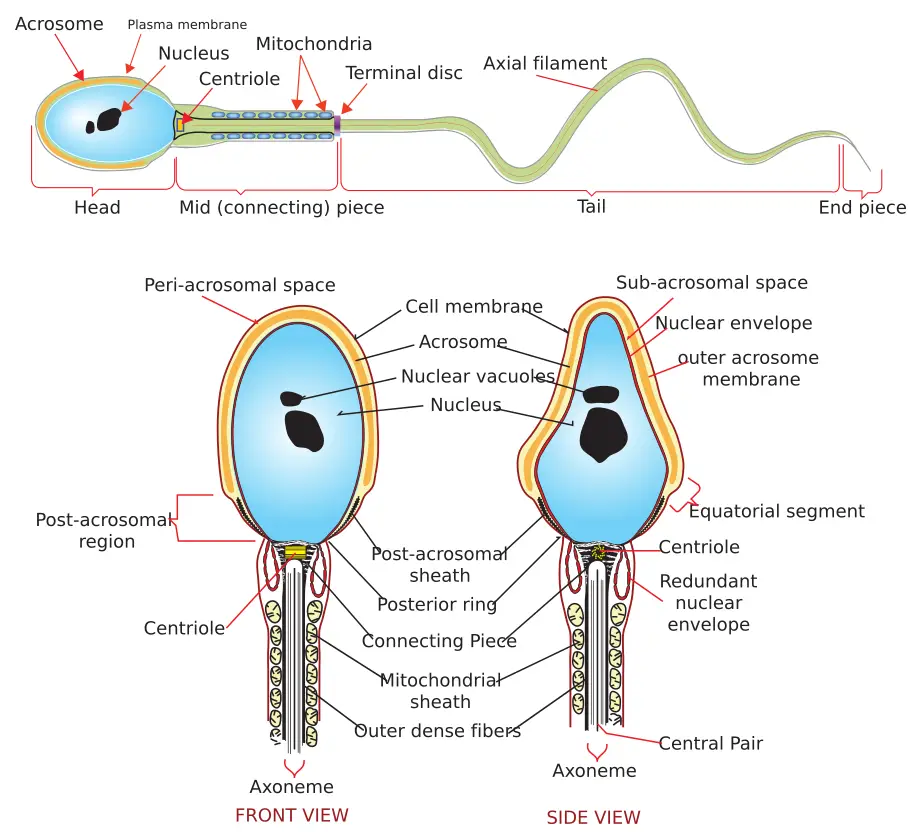
The primary reproductive organs in the male are the testes. Each testis is oval and measures about 1.6 inches (4 cm) long and 1 inch (2.5 cm) wide. The testes are surrounded by a white, fibrous capsule and are suspended together in a sac outside the body called the scrotum. Testes produce the gametes called spermatozoa. Spermatozoa are made up of three parts:

  • Head: Contains the chromosomes in the nucleus; the acrosome covers the head of the sperm and contains enzymes to help penetrate the ovum.
  • Midpiece: Contains mitochondria, which produce energy for movement.
  • Tail or flagellum: Used for movement.

Spermatogenesis

The formation of sperm is called spermatogenesis. The spermatozoa are formed in a series of tightly coiled tiny tubes in each testis called the seminiferous tubules. From the seminiferous tubules, the formed spermatozoa travel to the epididymis, where they mature and are stored. The epididymis is a coiled tube almost 20 feet (6 meters) long and rests on top of and behind each testis.

Structural parts of a sperm cell labeled
Parts of a sperm
Wikimedia Commons
/
Public Domain

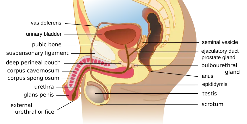
From the epididymis, the spermatozoa move into the vas deferens or ductus deferens. Each vas deferens is a muscular tunnel about 18 inches (45 cm) long that connects to the base of the epididymis and passes along the side of the testis. The vas deferens travels into the pelvic cavity to just behind the bladder. Spermatozoa can stay in the vasa deferentia for several months in an inactive state.

The prostate gland is the size of a walnut and is found below the bladder in males. It surrounds part of the urethra. The prostate is considered a reproductive organ but it also affects the urinary system. When the prostate increases in size (as with benign prostatic hyperplasia [BPH]), it can obstruct the urethra, blocking the urine flow.

The prostate gland, seminal vesicles, and Cowper glands (or bulbourethral glands) provide fluid either to nourish or aid in motility and lubrication. The sperm and the fluid together make up a substance called semen. The ejaculatory duct begins where the seminal vesicles join the vas deferens, and this “tube” joins the urethra.

Urethra and penis

The urethra is found within the penis and transports the semen to the outside of the body. The urethra also transports urine to the outside of the body.

The body is the shaft of the penis. The penis is composed of three columns of highly vascular erectile tissue. There are two columns of corpora cavernosa and one of corpus spongiosum that fill with blood through the dorsal veins during sexual arousal. During ejaculation, the sperm exit through the glans penis, the tip of the penis. At birth, the glans penis is surrounded by a fold of skin called the prepuce or foreskin, which can be removed by circumcision.

Physiology of the male reproductive system

Several hormones are involved in the male reproductive system, including testosterone, follicle-stimulating, and luteinizing hormones.

Organs of the male reproductive system labeled
The male reproductive system
Wikimedia Commons
/
CC BY-SA 4.0

Testosterone

At puberty , the interstitial cells in the testicles begin to produce testosterone . Testosterone is responsible for maintaining reproductive structures and for the development of sperm cells. Testosterone also helps with the development of secondary sex characteristics, which include a deep voice, broad shoulders, narrow hips, and additional body hair.

Follicle-stimulating and luteinizing hormones

In addition to testosterone, follicle-stimulating hormone (FSH), secreted by the pituitary gland, promotes the formation of spermatozoa. The pituitary gland also produces luteinizing hormone (LH), which stimulates the interstitial cells to produce testosterone. Under the influence of these hormones, sperm cells develop and move into the epididymis to mature.

Life span changes of the male reproductive system

The male reproductive system changes with age. The following sections discuss the changes to the male reproductive system over the life span.

Changes with puberty

The testicle develops in the abdomen and starts its descent into the scrotum in utero around the seventh month. At puberty, usually between ages 9 to 15, the pituitary gland secretes luteinizing hormone, which stimulates the testicles to start producing testosterone. Testosterone is responsible for the development of sperm and the secondary sex characteristics. The male secondary sex characteristics include a deep voice, broad shoulders, narrow hips, and additional body hair.

Changes in older adults

As a male gets older, gradual aging changes begin. Changes occur in the testicular tissue, sperm production, and erectile function. The testicular tissue mass decreases. Testosterone levels gradually decrease and may cause issues with erections. The testes continue to create sperm but at a slower rate. Benign prostatic hyperplasia can occur, leading to issues with urine retention, ejaculation, and a slowed urine stream.

Anatomy of the male reproductive system

  • Primary organs: testes (gonads), produce sperm and hormones
  • Sperm structure: head (chromosomes, acrosome), midpiece (mitochondria), tail (movement)
  • Accessory organs: epididymis (maturation/storage), vas deferens (transport), prostate/seminal vesicles/Cowper glands (fluid for semen), penis (urethra for semen/urine)

Physiology of the male reproductive system

  • Testosterone: produced by interstitial cells, develops/maintains reproductive structures and secondary sex characteristics
  • FSH: stimulates sperm production in testes
  • LH: stimulates testosterone production

Life span changes of the male reproductive system

  • Puberty: testicles descend, testosterone increases, secondary sex characteristics develop
  • Older adults: decreased testicular mass, lower testosterone, reduced sperm production, increased risk of BPH

Male reproductive system disorders

  • Common symptoms: redness, swelling, lumps, urinary/sexual dysfunction

Cancers of the male reproductive system

  • Prostate cancer: most common, risk factors include age, family history, African American ethnicity, high calcium, obesity
    • Symptoms: urinary issues, hematuria, erectile dysfunction, pain
    • Diagnosis: DRE, PSA blood test, biopsy, imaging
  • Testicular cancer: most common in ages 15-35, highly treatable
    • Risk factors: abnormal development, undescended testicle, family history
    • Diagnosis: ultrasound, CT, biopsy, tumor markers (AFP, beta-hCG, LDH)

Prostate-specific antigen blood test

  • PSA: protein made by prostate, elevated in cancer, BPH, prostatitis
  • Used for screening, not definitive for cancer diagnosis

Congenital male reproductive system diseases and disorders

  • Anorchism: absence of testes
  • Chordee: downward penile curve
  • Cryptorchidism: undescended testicle, treated with hormones/surgery
  • Epispadias: urethra opens on top of penis
  • Hypospadias: urethra opens on underside of penis
  • Hydrocele: fluid-filled sac in scrotum
  • Phimosis: tight foreskin, may block urethra

Inflammatory male reproductive system diseases and disorders

  • Balanitis: inflammation of glans/foreskin
  • Epididymitis: inflammation of epididymis, often bacterial
  • Orchitis: testicular inflammation, often viral/bacterial
  • Prostatitis: inflammation of prostate, urinary obstruction

Structural and functional male reproductive system diseases and disorders

  • Benign prostatic hyperplasia (BPH): enlarged prostate, urinary symptoms
    • Diagnosis: DRE, PSA, urinalysis, imaging
    • Treatment: lifestyle, alpha-blockers, 5-alpha reductase inhibitors, TURP, laser therapy
  • Erectile dysfunction (ED): inability to maintain erection, multiple causes (vascular, psychological, neurological)
    • Treatment: lifestyle changes, oral medications (Viagra, Cialis, etc.)
  • Male infertility: inability to conceive after 1 year, causes include varicocele, hormone imbalance, blockages, toxins
    • Diagnosis: semen analysis, hormone testing, imaging
    • Treatment: surgery, antibiotics, hormones, ART

Additional structural and functional male reproductive disorders

  • Peyronie’s disease: penile curvature from scar tissue
  • Priapism: persistent, painful erection
  • Gynecomastia: male breast tissue enlargement
  • Spermatocele: cyst in epididymis with fluid/dead sperm
  • Testicular torsion: twisting of spermatic cord, blocks blood supply
  • Varicocele: enlarged scrotal veins, can cause infertility

Sign up for free to take 15 quiz questions on this topic

Previous
Next  | 18.5 Medical assistant's role in urinary examinations and treatments
All rights reserved ©2016 - 2026 Achievable, Inc.

Male reproductive system anatomy and physiology

Anatomy of the male reproductive system

The male reproductive system is responsible for producing, transporting, and sustaining sperm cells. The sperm cells are called sex cells or gametes. A sperm cell contributes half of the genes of the new offspring. The gametes are produced in organs called gonads. Gonads are considered primary or essential sex organs. They are also responsible for producing hormones. The other organs in the reproductive system are secondary or accessory organs.

Testis

The primary reproductive organs in the male are the testes. Each testis is oval and measures about 1.6 inches (4 cm) long and 1 inch (2.5 cm) wide. The testes are surrounded by a white, fibrous capsule and are suspended together in a sac outside the body called the scrotum. Testes produce the gametes called spermatozoa. Spermatozoa are made up of three parts:

  • Head: Contains the chromosomes in the nucleus; the acrosome covers the head of the sperm and contains enzymes to help penetrate the ovum.
  • Midpiece: Contains mitochondria, which produce energy for movement.
  • Tail or flagellum: Used for movement.

Spermatogenesis

The formation of sperm is called spermatogenesis. The spermatozoa are formed in a series of tightly coiled tiny tubes in each testis called the seminiferous tubules. From the seminiferous tubules, the formed spermatozoa travel to the epididymis, where they mature and are stored. The epididymis is a coiled tube almost 20 feet (6 meters) long and rests on top of and behind each testis.

From the epididymis, the spermatozoa move into the vas deferens or ductus deferens. Each vas deferens is a muscular tunnel about 18 inches (45 cm) long that connects to the base of the epididymis and passes along the side of the testis. The vas deferens travels into the pelvic cavity to just behind the bladder. Spermatozoa can stay in the vasa deferentia for several months in an inactive state.

The prostate gland is the size of a walnut and is found below the bladder in males. It surrounds part of the urethra. The prostate is considered a reproductive organ but it also affects the urinary system. When the prostate increases in size (as with benign prostatic hyperplasia [BPH]), it can obstruct the urethra, blocking the urine flow.

The prostate gland, seminal vesicles, and Cowper glands (or bulbourethral glands) provide fluid either to nourish or aid in motility and lubrication. The sperm and the fluid together make up a substance called semen. The ejaculatory duct begins where the seminal vesicles join the vas deferens, and this “tube” joins the urethra.

Urethra and penis

The urethra is found within the penis and transports the semen to the outside of the body. The urethra also transports urine to the outside of the body.

The body is the shaft of the penis. The penis is composed of three columns of highly vascular erectile tissue. There are two columns of corpora cavernosa and one of corpus spongiosum that fill with blood through the dorsal veins during sexual arousal. During ejaculation, the sperm exit through the glans penis, the tip of the penis. At birth, the glans penis is surrounded by a fold of skin called the prepuce or foreskin, which can be removed by circumcision.

Physiology of the male reproductive system

Several hormones are involved in the male reproductive system, including testosterone, follicle-stimulating, and luteinizing hormones.

Testosterone

At puberty , the interstitial cells in the testicles begin to produce testosterone . Testosterone is responsible for maintaining reproductive structures and for the development of sperm cells. Testosterone also helps with the development of secondary sex characteristics, which include a deep voice, broad shoulders, narrow hips, and additional body hair.

Follicle-stimulating and luteinizing hormones

In addition to testosterone, follicle-stimulating hormone (FSH), secreted by the pituitary gland, promotes the formation of spermatozoa. The pituitary gland also produces luteinizing hormone (LH), which stimulates the interstitial cells to produce testosterone. Under the influence of these hormones, sperm cells develop and move into the epididymis to mature.

Life span changes of the male reproductive system

The male reproductive system changes with age. The following sections discuss the changes to the male reproductive system over the life span.

Changes with puberty

The testicle develops in the abdomen and starts its descent into the scrotum in utero around the seventh month. At puberty, usually between ages 9 to 15, the pituitary gland secretes luteinizing hormone, which stimulates the testicles to start producing testosterone. Testosterone is responsible for the development of sperm and the secondary sex characteristics. The male secondary sex characteristics include a deep voice, broad shoulders, narrow hips, and additional body hair.

Changes in older adults

As a male gets older, gradual aging changes begin. Changes occur in the testicular tissue, sperm production, and erectile function. The testicular tissue mass decreases. Testosterone levels gradually decrease and may cause issues with erections. The testes continue to create sperm but at a slower rate. Benign prostatic hyperplasia can occur, leading to issues with urine retention, ejaculation, and a slowed urine stream.

Key points

Anatomy of the male reproductive system

  • Primary organs: testes (gonads), produce sperm and hormones
  • Sperm structure: head (chromosomes, acrosome), midpiece (mitochondria), tail (movement)
  • Accessory organs: epididymis (maturation/storage), vas deferens (transport), prostate/seminal vesicles/Cowper glands (fluid for semen), penis (urethra for semen/urine)

Physiology of the male reproductive system

  • Testosterone: produced by interstitial cells, develops/maintains reproductive structures and secondary sex characteristics
  • FSH: stimulates sperm production in testes
  • LH: stimulates testosterone production

Life span changes of the male reproductive system

  • Puberty: testicles descend, testosterone increases, secondary sex characteristics develop
  • Older adults: decreased testicular mass, lower testosterone, reduced sperm production, increased risk of BPH

Male reproductive system disorders

  • Common symptoms: redness, swelling, lumps, urinary/sexual dysfunction

Cancers of the male reproductive system

  • Prostate cancer: most common, risk factors include age, family history, African American ethnicity, high calcium, obesity
    • Symptoms: urinary issues, hematuria, erectile dysfunction, pain
    • Diagnosis: DRE, PSA blood test, biopsy, imaging
  • Testicular cancer: most common in ages 15-35, highly treatable
    • Risk factors: abnormal development, undescended testicle, family history
    • Diagnosis: ultrasound, CT, biopsy, tumor markers (AFP, beta-hCG, LDH)

Prostate-specific antigen blood test

  • PSA: protein made by prostate, elevated in cancer, BPH, prostatitis
  • Used for screening, not definitive for cancer diagnosis

Congenital male reproductive system diseases and disorders

  • Anorchism: absence of testes
  • Chordee: downward penile curve
  • Cryptorchidism: undescended testicle, treated with hormones/surgery
  • Epispadias: urethra opens on top of penis
  • Hypospadias: urethra opens on underside of penis
  • Hydrocele: fluid-filled sac in scrotum
  • Phimosis: tight foreskin, may block urethra

Inflammatory male reproductive system diseases and disorders

  • Balanitis: inflammation of glans/foreskin
  • Epididymitis: inflammation of epididymis, often bacterial
  • Orchitis: testicular inflammation, often viral/bacterial
  • Prostatitis: inflammation of prostate, urinary obstruction

Structural and functional male reproductive system diseases and disorders

  • Benign prostatic hyperplasia (BPH): enlarged prostate, urinary symptoms
    • Diagnosis: DRE, PSA, urinalysis, imaging
    • Treatment: lifestyle, alpha-blockers, 5-alpha reductase inhibitors, TURP, laser therapy
  • Erectile dysfunction (ED): inability to maintain erection, multiple causes (vascular, psychological, neurological)
    • Treatment: lifestyle changes, oral medications (Viagra, Cialis, etc.)
  • Male infertility: inability to conceive after 1 year, causes include varicocele, hormone imbalance, blockages, toxins
    • Diagnosis: semen analysis, hormone testing, imaging
    • Treatment: surgery, antibiotics, hormones, ART

Additional structural and functional male reproductive disorders

  • Peyronie’s disease: penile curvature from scar tissue
  • Priapism: persistent, painful erection
  • Gynecomastia: male breast tissue enlargement
  • Spermatocele: cyst in epididymis with fluid/dead sperm
  • Testicular torsion: twisting of spermatic cord, blocks blood supply
  • Varicocele: enlarged scrotal veins, can cause infertility

More from Anatomy and physiology of the urinary system

  • Urinary system anatomy
  • Urinary system diseases and disorders
  • Chronic kidney disease and nephrotic syndrome
  • Medical assistant's role in urinary examinations and treatments