



Environmental and traumatic skin disorders: burns, cold injuries, and carcinomas
Burns
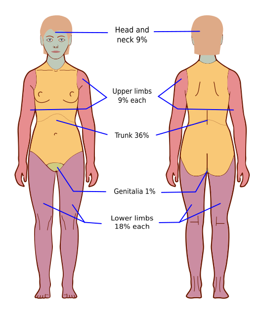
Burns are injuries to tissues that result from exposure to thermal, chemical, electrical, or radioactive agents. They are classified into four different degrees of severity, depending on the layers of the skin that are damaged. Burns that are second degree or higher should be further categorized according to the “rule of nines.” The rule of nines regarding burns divides the body into percentages that are, for the most part, multiples of nine: the head and neck equal 9%, each upper limb 9%, each lower limb 18%, the front of the torso 18%, back of the torso 18%, and the genital area 1%.
Because burns are injuries, the cause is trauma of some kind. Many risk factors could be included in this discussion, but particular caution should be exercised when participating in activities that involve fire, chemicals, or electrical hazards. Proper safety equipment should always be available and ready to use when burns are a possibility.
The categories of burns are as follows:
- First-degree burn (superficial burn): A burn in which only the first layer of the skin, the epidermis, is damaged. It is characterized by redness (erythema), tenderness, and physical sensitivity (hyperesthesia), with no scar development.
- Second-degree burn (partial-thickness burn): A burn in which only the first and second layers of the skin (the epidermis and part of the dermis) are affected. If the burn extends to the papillary level, it is classified as a superficial partial-thickness burn. If it extends farther to the reticular layer, it is classified as a deep partial-thickness burn. This type of burn is characterized by redness, blisters, and pain, with possible scar development.
- Third-degree burn (full-thickness burn): A burn that damages the epidermis, dermis, and subcutaneous tissue. Pain is not present because the nerve endings in the skin have been destroyed. The skin may appear deep red, pale gray, brown, or black. Scar formation is likely.
- Fourth-degree burn (deep full-thickness burn): Although not a universally accepted category, some burn specialists use this category to describe a rare burn that extends beyond the subcutaneous tissue into the muscle and bone.
Burns may not uniformly affect an injured area, so determining the depth of a burn and the extent of tissue damage is important. Burns are treated differently depending on the depth of damage. Treatment is based on the category of burn.
The American Burn Association defines a severe burn as a burn that affects 25% of the total body surface area or any burn involving the eyes, ears, face, hands, feet, or groin. With a severe burn, the patient may require additional testing, such as laboratory tests, x-rays, or other diagnostics.
Cold injuries
Cold injuries usually are less severe than burns, but prolonged exposure to cold temperatures can result in infection, gangrene, amputation and, in severe cases, death. Frostbite is caused by exposure to subfreezing temperatures. Damage occurs at the level of the capillaries, which become permanently dilated and unable to regulate local blood flow. Signs and symptoms of superficial frostbite include burning, tingling, numbness, and a white or grayish color of the skin. With deep frostbite, blisters form, and the area is hard, mottled, edematous, and blue or gray after thawing.
Carcinomas of the skin
Skin cancer is the abnormal, malignant growth of skin cells. Skin that is exposed to the sun is at higher risk for developing skin cancer, but skin cancer can also occur on skin not ordinarily exposed to sunlight.
There are three major types of skin cancer:
- Basal cell carcinoma
- Squamous cell carcinoma
- Melanoma, often called malignant melanoma
Skin cancer starts with mutations that occur in the DNA of skin cells. The mutations cause the cells to grow out of control and form cancer cells. Skin cancer begins in the epidermis.
Skin cancer can happen in three different types of cells in the epidermis:
- Squamous cells lie just below the outer surface of the skin.
- Basal cells produce new skin cells and are located beneath the squamous cells.
- Melanocytes produce melanin, the pigment that gives skin its normal color. They are located in the lower part of the epidermis.
These cancers are thought to be caused by exposure to UV rays of the sun. UV rays can damage DNA in the skin’s cells, which starts a mutation. However, not all cancers develop in areas of the body exposed to the sun; it is still unknown what other specific factors influence the start of a malignant mutation. Additional risk factors for skin cancer include fair skin with blond or red hair, light-colored eyes, and skin that freckles or sunburns easily; a history of sunburns that have blistered; moles that look irregular and are generally larger than normal moles; and a family history of skin cancer (if one parent or a sibling has had skin cancer, there may be an increased risk of the disease).
Basal cell carcinoma
Basal cell carcinoma most frequently appears on sun-exposed areas (FIGURE 17.17) and may appear as follows:
- A pearly or waxy bump
- A flat, flesh-colored or brown scar like lesion
Squamous cell carcinoma
Squamous cell carcinoma most frequently appears on sun-exposed areas and may appear as follows:
- A firm, red nodule
- A flat lesion with a scaly, crusted surface
Melanoma
Melanoma can develop anywhere on the body. It can start in normal-looking skin or an existing mole. Skin that has not been exposed to sunlight can still develop melanoma. Melanoma can affect people of any skin color. People with darker skin can develop melanoma on the palms of their hands or the soles of their feet, or even under fingernails or toenails. Melanoma may appear as follows:
- A large brownish spot with darker speckles
- A mole that changes in color, size, or feel or that bleeds
- A small lesion with an irregular border and portions that appear red, white, blue, or blue-black
- Dark lesions on the palms of the hands, soles of the feet, fingertips, or toes
To diagnose any type of skin cancer, the provider will do the following:
- Examine the skin and look for changes that are likely to be skin cancer.
- If abnormal areas of the skin are found, a skin biopsy will be performed. A biopsy will determine whether skin cancer is present and what type of cancer it is.
Additional tests may be done to determine the extent or stage of the skin cancer. These tests might include the following:
- Imaging tests to examine nearby lymph nodes for signs of cancer.
- Removal of a nearby lymph node that will be tested for signs of cancer and staged; a skin cancer’s stage helps determine which treatment options will be most effective.
Treatment for skin cancer varies greatly, depending on the type of skin cancer and its stage. Early detection and treatment are always the best options for any type of skin cancer, but that is not always possible.
Common treatments for basal cell and squamous cell carcinomas include the following:
- Mohs micrographic surgery
- Excisional surgery
- Topical medications and photodynamic therapy (PDT)
- Electrosurgery, cryosurgery, or laser surgery
Treatment for melanoma varies greatly, depending on the location and stage of the cancer. Treatment ranges from minor localized surgery to major surgery, radiation, and chemotherapy.