



Assisting with the nervous system
The anatomy and physiology of the nervous system
The nervous system works with the endocrine system to integrate stimuli, both from within the body and from the outside environment, to regulate body systems; this allows homeostasis to be maintained. The nervous system is divided into two major parts: the central nervous system (CNS), which is made up of the brain and spinal cord, and the peripheral nervous system (PNS), which includes all the nervous tissue and neurologic responses found outside the CNS.
The brain is the “president” or “chief executive officer” of the body. It constantly receives information from the periphery, including all the organs and systems inside the body and on its surface. This information (i.e., stimuli) is carried to the brain by the peripheral nerves along the afferent, or ascending, nerve tract. The brain monitors and interprets the stimuli received from the afferent nerves and sends appropriate responses back along efferent pathways to the organs or to the body’s surface. These responses from the brain cause a specific reaction in the organ, in the glands, or in skeletal muscles. These reactions keep the body running smoothly and allow it to react instantly to both external and internal stimuli.
The functioning cell of the nervous system is the neuron. The brain contains billions of individual neurons. Formation of the nervous system starts very early in embryonic development (i.e., by week 3); it begins as the neural tube, which eventually develops into the brain and spinal cord. Each neuron is made up of a main cell body that contains the nucleus and a relatively long extension of the cell, called the axon, which may be covered with a myelin sheath. Multiple filaments, called dendrites, extend from the neuron body. Dendrites receive the nervous impulse from a preceding neuron and carry it into the cell body. Impulses are carried away from the cell body through the axon to another neuron or to cells in another tissue. This transfer of stimuli begins as an electrical impulse that travels down an axon of one neuron and becomes a chemical impulse while moving across the synapse (the space between two neurons) to the dendrite of another neuron. The transfer of impulses from the end of one neuron to the dendrites of another is enhanced by chemical neurotransmitters, which bind to specific receptor sites on the dendrites of the next neuron. If the nerve impulse is traveling to a muscle or to any other organ or tissue instead of another neuron, the chemical neurotransmitters bind to special receptors in the target tissue. Messages move throughout the entire nervous system in this manner. Impulses in the neuron are electrical; the impulses become chemical as a specific neurotransmitter is released at each synapse, and they become electrical again as they are picked up by the subsequent dendrites of another neuron or by the target tissue.
Supportive cells of the nervous system are called glial or neuroglial cells. Glial cells do not carry on any of the functions of the nervous system; these specialized cells perform specific functions within the nervous system; for example, Schwann cells form the myelin sheath, and astrocytes help form the blood-brain barrier. The blood-brain barrier closely regulates what substances enter the brain tissue. Oxygen, water, and glucose molecules easily pass into the brain, whereas many chemicals and drugs are prevented from moving into brain tissue. Brain inflammation can increase the ability of many drugs to cross the blood-brain barrier because of damage to the specialized glial cells.
Central nervous system
As mentioned, the brain and spinal cord together make up the CNS. The brain is encased within the skull in the cranial cavity. The spinal cord is a bundle of nervous tissue that extends inferiorly from the brainstem at the base of the brain and exits the skull at the foramen magnum. It descends for about 17 inches inside the spinal canal, which courses through the vertebrae of the backbone.
Brain
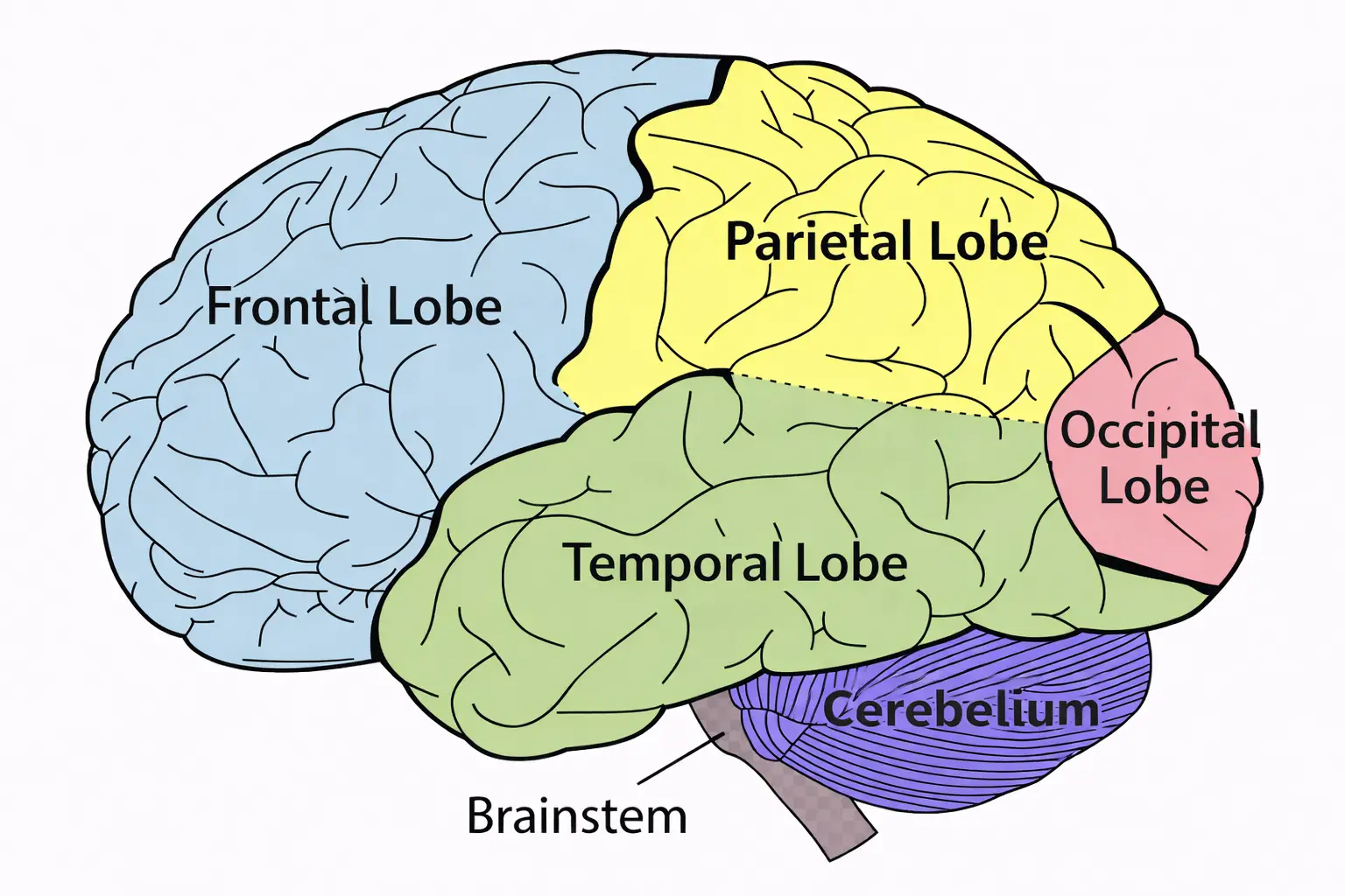
The brain accounts for only about 2% of a person’s weight, but it consumes about 20% of the body’s oxygen. The brain is divided into three main areas: the cerebrum, the cerebellum, and the brainstem. The cerebrum, the largest and uppermost section of the brain, has multiple convolutions along its surface, called gyri, which are formed by the folding in of the cerebral cortex. The gyri are separated by shallow grooves, called sulci. The gyri greatly increase the surface area of the cerebrum, which maximizes the potential of the CNS neurons in each area. The cerebrum is divided into lobes, which are named after the region of the skull under which they are located. The cerebrum is separated by a longitudinal fissure into left and right hemispheres. The right hemisphere usually controls artistic functions, such as drawing, rhythm, and picture memory. The left hemisphere controls verbal functions, such as reading, writing, speaking, and mathematic calculations. The two halves of the brain are connected by the corpus callosum. This bundle of nerve tissue facilitates communication between the two sides of the brain. The corpus callosum is the largest collection of white matter within the brain; it has a high myelin content, which allows for quicker transmission of information. Some congenital defects include a complete lack of this neural tissue.
The diencephalon, located deep in the center of the cerebrum near the superior portion of the brainstem, is made up of the thalamus and the hypothalamus. The thalamus acts as a relay station between sensory neurons and the cerebral cortex. The functions of the hypothalamus include controlling the autonomic nervous system; regulating endocrine processes; and managing body temperature, sleep, and appetite to maintain homeostasis. Within the cerebrum are four spaces, called ventricles, which contain cerebrospinal fluid (CSF). CSF nourishes, lubricates, and provides some cushioning protection for the brain and the spinal cord.
The cerebellum, which is just inferior to the occipital lobe of the cerebrum, controls balance, equilibrium, posture, and muscle coordination. The brainstem controls reflexes and serves as a sensory relay station for input coming into the brain from the body. The brainstem plays a vital role in vision, hearing, respiration, heart rate, blood pressure, waking, and sleeping.
Spinal cord
The spinal cord extends from the inferior portion of the brainstem to approximately the second lumbar vertebra. Thirty-one pairs of spinal nerves extend from the spinal cord through openings in the vertebrae. Starting just below the first cervical vertebra in the neck, a nerve extends from the spinal cord on each side; therefore, a pair of spinal nerves originates at each level. Each of these pairs of nerves innervates a specific organ or area of the body. The spinal cord carries messages between the spinal nerves and the brain.
Meninges
Because the brain and the spinal cord are critical to life, they are well protected. They both are encased in some of the thickest bones in the body; they also are surrounded by three membranes, called meninges; and they are cushioned by the CSF. The outer layer of the meninges is called the dura mater (“hard mother”) because it is a tough membrane, similar to a very strong rubber band. The subdural space lies below the dura mater and contains small veins that have little protection. Trauma to the head can cause bleeding of these tiny vessels, ultimately leading to the development of a subdural hematoma. Above the dura mater is the epidural space. The arterial supply to the meninges comes from blood vessels that line the inner aspect of the skull. If the skull is fractured, these arteries can be damaged, resulting in a collection of blood between the skull and the dura mater called an epidural hematoma.
The middle meningeal layer is the arachnoid, which was given that name because of its fine spider-web appearance. Beneath the arachnoid membrane in the subarachnoid space is the cerebrospinal fluid, a clear liquid that contains glucose, protein, and chloride produced by specialized cells in the ventricles. CSF circulates continuously through the ventricles and around the brain and spinal cord, carrying nutrients and removing wastes. The innermost meningeal layer, which covers the brain and spinal cord, is the delicate pia mater (“tender mother”); it is highly vascular and the thinnest of the three layers. The pia mater provides support for the blood vessels of the brain.
Hydrocephalus
Hydrocephalus is the abnormal accumulation of cerebrospinal fluid (CSF) in the ventricles of the brain. It can be detected in utero with sonography or diagnosed at birth. It is the result either of overproduction of CSF or of failure of the fluid to drain properly. If left untreated, hydrocephalus causes gross enlargement of the skull and severe damage to brain tissue from increased intracranial pressure. The only treatment is surgery to place a shunt (tube) from a ventricle in the brain to the right atrium or to the abdominal cavity. The shunt allows the excess CSF to drain away from the brain.
Peripheral nervous system
The PNS is made up of the nerves that exit the brain or spinal cord. The peripheral nerves exiting the brain directly through the cranium are called cranial nerves. Cranial nerves originate from the underside of the brain and relay information to and from the sensory organs and muscles of the face and neck. The spinal nerves from the spinal cord enter and exit the spinal canal through spaces between the vertebrae. Spinal nerves carry information to and from the brain through the spinal cord. Sensory fibers in these nerves carry stimuli from the skin and internal organs to the CNS. Motor fibers carry messages from the CNS to skeletal muscles, causing them to contract. The autonomic nervous system (ANS) is part of the PNS. Autonomic nerves control homeostasis; that is, they keep the body running smoothly, much like a thermostat controls the temperature in a room. The ANS is an automatic system that regulates body functions such as breathing, heart rate, sweating, circulation, and digestion. It also controls the actions of muscles in blood vessel walls, organs, and glands. Just as a thermostat can control both heating and cooling in a room to maintain a comfortable temperature, the autonomic system is made up of two divisions, called the sympathetic system and the parasympathetic system. The sympathetic system promotes responses geared toward protecting the individual 522(“fight or flight”), generally causing a stimulating effect: it speeds up the heart, raises blood glucose levels and blood pressure, reduces peristalsis, and widens the bronchioles, allowing more oxygen to enter the body quickly. The parasympathetic system generally promotes rest or a reducing effect: it slows the heart rate, constricts the bronchioles, and increases digestive system function.