


Achievable logoAchievable logo
CCMA
Sign in
Sign up
Purchase
Textbook
Practice exams
Support
How it works
Exam catalog
Mountain with a flag at the peak
Textbook
1. Medical assistant
2. Electronic records
3. Medical terminology and anatomy
4. The fundamentals of infection control
5. Introduction to vital signs
6. The patient interview and history
7. The physical examination
8. Appointment scheduling
9. Insurance billing
10. Diagnostic coding and the ICD-10-CM System
11. Procedural coding
12. Medical billing and reimbursement essentials
13. Assisting with medical specialties
14. Assisting with the musculoskeletal system
15. Assisting with the cardiovascular system
16. Assisting with the respiratory system
16.1 Assisting with the respiratory system
16.2 Respiratory diseases: signs, symptoms, and chronic conditions
16.3 Common acute respiratory diseases
16.4 Tobacco use and other chronic respiratory disorders
16.5 Severe and additional acute respiratory disorders
16.6 Medical assistant's role in respiratory examinations and diagnostics
17. Assisting with the nervous system
18. Anatomy and physiology of the urinary system
19. Assisting in obstetrics and gynecology
20. Assisting in endocrinology
21. Assisting in ophthalmology & otolaryngology
22. Assisting in gastroenterology
23. Assisting in the immune & lymphatic systems
24. Assisting in pediatrics: the developmental stages and care
25. The medical assistant’s role in caring for the older patient
26. The role of the medical assistant in physical therapy examination and assessment
27. Preparing for minor surgery: room, solutions, and supplies
28. Introduction to the clinical laboratory
29. Urinalysis
30. Blood collection
31. Analysis of blood
32. Electrocardiography and heart structure
33. The principles of pharmacology
34. Essential calculations and measurement systems
35. Solid, liquid, & solutions medication doses
36. Administering medications
37. Metabolism and core nutrient roles
38. Medical emergencies in the healthcare setting
Achievable logoAchievable logo
16.1 Assisting with the respiratory system
Achievable CCMA
16. Assisting with the respiratory system
Our CCMA course is currently in development and is a work-in-progress.

Assisting with the respiratory system

10 min read
Font
Discuss
Share
Feedback

Pulmonology is the healthcare specialty that deals with respiratory diseases and disorders. A pulmonologist is a specialist involved in the diagnosis, treatment, and prevention of disorders of the respiratory system.

Pulmonary procedures and treatments are common in the ambulatory care area. Besides the pulmonary department, patients with respiratory concerns are typically seen in primary and urgent care departments. Medical assistants measure peak flow rates, perform spirometry, and assist with pulmonary treatments. Nebulizer treatments and oxygen therapy are the most frequent pulmonary treatments in ambulatory care.

The respiratory tract

The respiratory system is divided into the upper respiratory tract and the lower respiratory tract. The upper respiratory tract structures are considered passageways for the air, whereas the lower respiratory tract structures are involved in gas exchange.

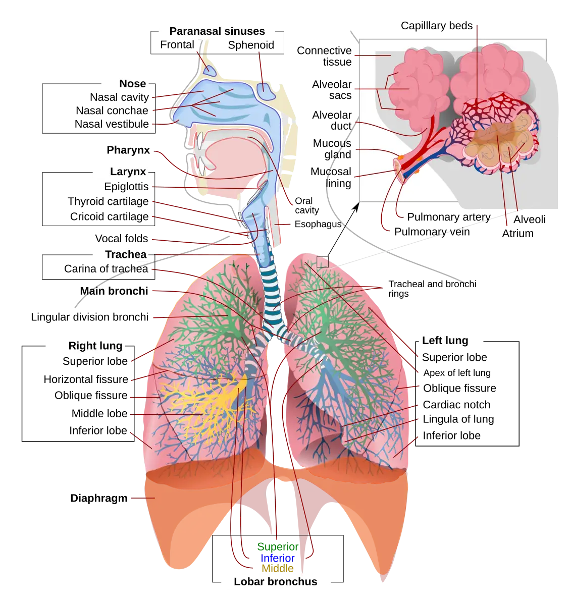
Structures of the respiratory system and lungs labeled
Respiratory system and lungs
Wikimedia Commons
/
Public Domain

The anatomy of the respiratory tract

Anatomy of the upper respiratory tract

The upper respiratory tract is composed of structures from the nose to the larynx. These organs are located outside the chest cavity. The upper respiratory tract has several functions:

  • Warming and cleaning the inspired air
  • Serving as a passageway for air
  • Providing the sense of smell

Nose and paranasal sinuses

Air can enter through the mouth or the two nares (nostrils) in the nose. The nasal septum separates the nares. The air then moves into the nasal cavity. The surface capillaries, mucous membrane, and cilia (small hairs) found in the nasal cavity clean, warm, and moisten the air. The cilia continually move in a wavelike motion to push mucus and debris out of the respiratory tract. Receptors for smell are in the nasal cavity. The nasal cavity is connected to four pairs of paranasal sinuses. The four pairs of paranasal sinuses are named for the bones they are found in: maxillary, frontal, sphenoid, and ethmoid.

Pharynx

Air continues to travel into the** pharynx (throat)**, which is divided into three sections:

  • Nasopharynx: Located behind the nasal cavity. The eustachian tube connects the middle ear to the nasopharynx and equalizes the pressure in the ear with the air pressure outside the body.
  • Oropharynx: Located behind the mouth and part of the respiratory and digestive systems.
  • Laryngopharynx: Located between the epiglottis and the esophagus.

The epiglottis, a flap of cartilage at the larynx opening, closes off the trachea when food is swallowed. As air passes out through the opening, the larynx (vocal cords) vibrates to produce speech. The vocal cords are paired bands of cartilaginous tissue.

Anatomy of the lower respiratory tract

The lower respiratory tract consists of the trachea, bronchial tubes, and lungs. These structures are lined with mucous membranes and cilia. The** trachea (windpipe)** lies in the space between the lungs, called the mediastinum. Air travels from the larynx through the trachea, and then the trachea branches into the right and left bronchi. The right bronchus is wider than the left bronchus.

Bronchioles and Alveoli

The bronchi divide into smaller branches, called bronchioles. These bronchioles end in microscopic ducts capped by air sacs, called alveoli. Each thin-walled alveolus is in contact with a blood capillary. This contact between the two structures allows the exchange of gases. It is at this point that oxygen (O2) from the inspired air moves across the one-cell membrane into the blood cells. Carbon dioxide (CO2) moves in the other direction, from the blood into the air to be expired. Each alveolus is coated with a substance called surfactant, which keeps it from collapsing. Without surfactant, the alveoli stick together during exhalation (breathing out) and deflate. Inhalation (breathing in) becomes more difficult, and less O2 can move into the bloodstream. This condition is life-threatening. Babies born before 37 to 39 weeks of gestation are at risk of not having enough surfactant. If time permits before delivery, steroids can be given to the mother to help mature the baby’s lungs.

Lungs

The bronchial tree and alveoli are the major structures in the right and left lungs. The lungs are soft and spongy because of the air sacs that make up most of their mass. They hang on the right and left sides of the chest, separated by the pericardial sac, which contains the heart. Each lung is composed of sections called lobes. The right lung consists of three lobes, whereas the left has only two lobes.

Because each lobe has its own bronchus and blood supply, the removal of one lobe (lobectomy) results in little or no damage to the rest of the lung. The left lung is longer and narrower. It has a distinct indentation in its center, known as the cardiac notch. This is where the left ventricle of the heart is located and where an apical pulse is heard.

Pleura and pleural cavity

Pleura is a thin serous membrane found in the thoracic cavity. The pleura folds back on itself, creating a sac that surrounds the lung. The visceral pleura covers the lungs and adjoining structures. The parietal pleura, the outer portion of the pleura, lines the thoracic cavity and covers the diaphragm and mediastinum. Only the parietal pleura contains pain receptors, making it highly sensitive to pain. The right and left pleural sacs are entirely separate.

The pleural cavity is the space between the visceral and parietal pleurae. A small amount of pleural fluid is in the pleural cavity. When the lungs expand, moving the visceral pleura closer to the parietal pleura, friction is reduced between the tissues due to the pleural fluid.

Respiratory muscles

The muscles responsible for normal, quiet respiration are the diaphragm and the intercostal muscles. On inspiration, the diaphragm is pulled down and flattened as it contracts, and the intercostal muscles expand, pulling air into the lungs. On expiration, the diaphragm relaxes and moves upward, pushing air out of the lungs.

The mechanisms of breathing

The two primary functions of the respiratory system are to exchange O2 from the atmosphere for CO2 waste and to maintain the acid-base balance in the body. Both functions involve ventilation (breathing), which is the movement of gases between the lungs and the environment. Ventilation includes the process of inspiration and expiration.

Two types of respiration occur during the ventilation process:

  • External respiration occurs when oxygenated air moves into the alveoli. Surrounding each alveolus is a pulmonary capillary network. The alveoli and the pulmonary capillaries are made of single-celled walls. This allows the O2 to move easily across the alveoli and capillaries into the blood. CO2 and other wastes are forced out of the capillaries and move into the alveoli.
  • Internal respiration occurs when O2 is exchanged for CO2 between the cells in the body and the blood.

Inspiration

A healthy person breathes when the blood CO2 level increases. A person with** chronic obstructive pulmonary disease (COPD)** has a constantly elevated blood CO2 level. At some point, the body no longer uses the elevated blood CO2 level as a trigger to breathe. A secondary system kicks in, and breathing is triggered by a decreased blood O2 level.

When the breathing trigger is activated, it signals the respiratory center in the medulla oblongata, which is in the brainstem. The respiratory center causes a stimulus (i.e., signal) to be carried by the phrenic nerve to the diaphragm. When the diaphragm receives the signal, it flattens out and pulls downward. At the same moment, the intercostal muscles between the ribs contract, causing the ribs to move outward. The movement enlarges the chest cavity and causes the lungs to expand, increasing their volume. The greater the contraction, the deeper the inhalation and the greater the air volume.

Respiratory distress changes

When individuals are experiencing respiratory distress, they are unable to move enough air into the lungs. To help move additional air into their lungs, they use accessory muscles . To identify whether a person is using the accessory muscles, expose the chest and look for chest retractions with breathing. With intercostal retractions, the chest tissue between the ribs is indrawn or pulled in during breathing. Upper airway obstructions can cause the following retractions:

  • Suprasternal retractions, sucking in of the skin just above the sternum
  • Supraclavicular retractions, sucking in of the skin just above the clavicle
  • Lower airway obstructions can cause the following retractions:
  • Substernal retractions, sucking in of the abdomen just below the sternum
  • Subcostal retractions, sucking in of the abdomen just below the ribs

Using accessory muscles to breathe can tire a person. This can lead to** respiratory arrest** , a medical emergency. Typically, this occurs quicker in infants and young children.

Expiration

The second half of ventilation is expiration. Once inspiration is complete, the diaphragm and intercostal muscles relax, which causes the diaphragm to move upward into the thoracic cavity and the ribs to move inward. This movement reduces the lung capacity and forces air out of the lungs. Typically, expiration requires very little energy. However, with some conditions (e.g., asthma and emphysema), the person has difficulty getting air out of the lungs. The accessory muscles are needed to help with complete exhalation.

Acid-base balance

The body attempts to keep the pH between 7.35 and 7.45. The respiratory system has an important role in acid-base balance. It regulates the amount of CO2 in the blood. CO2 in the blood can combine with water to form the buffer bicarbonate. If a person hyperventilates (breathes rapidly), the CO2 and bicarbonate levels in the blood decrease. This causes the pH of the body to rise, resulting in respiratory alkalosis. This condition can be seen in patients with anxiety or an acute asthma attack. If hypoventilation occurs, the CO2 in the blood increases (hypercapnia), and respiratory acidosis can occur. Respiratory alkalosis and respiratory acidosis are both life-threatening disorders if the underlying causes are not corrected.

Life span changes

Changes occur in the respiratory system over a person’s lifetime. The following sections explain the changes that occur from infancy to adulthood.

Infant

An infant has a narrow airway with a shorter and softer trachea. If the neck is overextended, the airway can collapse. Infants tend to breathe through their noses, which means nasal congestion can make breathing difficult. Infants are abdominal breathers and have immature respiratory muscles, meaning fatigue with breathing difficulties can set in quickly. With a disproportionately larger tongue and epiglottis, infants and young children are at risk for airway obstruction.

Adult

At around age 20 to 25, the lungs reach maturity. By age 35, lung function starts to decline. People who smoke can increase the aging of their lungs. As a person ages, the following respiratory system changes occur:

  • Chest wall and thoracic spine deformities cause increased work of breathing.
  • The diaphragm grows weaker, leading to a decreased ability to inhale and exhale.
  • Weakness in the respiratory muscles causes the coughing reflex to be less effective.
  • A decrease in tissue elasticity leads to an inability to keep the airway completely open.
  • Alveoli lose their shape, which causes air to be trapped in the lungs. This leads to a decrease in gas exchange and lung capacity.

Pulmonology Overview

  • Specialty for respiratory diseases/disorders
  • Pulmonologists diagnose, treat, and prevent respiratory conditions
  • Common ambulatory procedures: peak flow, spirometry, nebulizer, oxygen therapy

Anatomy of the Upper Respiratory Tract

  • Structures: nose, nasal cavity, paranasal sinuses, pharynx, larynx
  • Functions: warms/cleans air, passageway, sense of smell
  • Pharynx sections: nasopharynx, oropharynx, laryngopharynx
    • Epiglottis prevents food entry into trachea
    • Larynx (vocal cords) produces speech

Anatomy of the Lower Respiratory Tract

  • Structures: trachea, bronchi, bronchioles, alveoli, lungs
  • Trachea divides into right (wider) and left bronchi
  • Bronchioles end in alveoli (site of gas exchange)
    • Alveoli coated with surfactant to prevent collapse

Lungs

  • Right lung: 3 lobes; left lung: 2 lobes (cardiac notch present)
  • Lobes have separate bronchi and blood supply
  • Lungs separated by pericardial sac (contains heart)

Pleura and Pleural Cavity

  • Pleura: serous membrane with two layers
    • Visceral pleura covers lungs; parietal pleura lines thoracic cavity
    • Only parietal pleura has pain receptors
  • Pleural cavity: space between pleurae, contains lubricating fluid

Respiratory Muscles

  • Diaphragm and intercostal muscles drive normal breathing
  • Inspiration: diaphragm contracts (moves down), intercostals expand chest
  • Expiration: diaphragm relaxes (moves up), air pushed out

Mechanisms of Breathing

  • Main functions: O2/CO2 exchange, acid-base balance
  • Ventilation = inspiration + expiration
  • Two types of respiration:
    • External: alveoli ↔ pulmonary capillaries
    • Internal: blood ↔ body cells

Inspiration

  • Triggered by increased blood CO2 (except in COPD—triggered by low O2)
  • Medulla oblongata (respiratory center) sends signal via phrenic nerve
  • Diaphragm and intercostals contract, expanding chest cavity

Respiratory Distress Changes

  • Use of accessory muscles indicates distress
    • Retractions: suprasternal, supraclavicular (upper airway); substernal, subcostal (lower airway)
  • Prolonged use leads to fatigue, risk of respiratory arrest (especially in infants/children)

Expiration

  • Diaphragm/intercostals relax, reducing lung volume, air exits
  • Normally passive; in asthma/emphysema, accessory muscles needed

Acid-Base Balance

  • Normal blood pH: 7.35–7.45
  • Respiratory system regulates CO2 (forms bicarbonate buffer)
  • Hyperventilation: ↓CO2, ↑pH (respiratory alkalosis)
  • Hypoventilation: ↑CO2, ↓pH (respiratory acidosis)
  • Both alkalosis and acidosis are life-threatening if untreated

Life Span Changes

  • Infant:
    • Narrow, soft airway; risk of collapse with neck extension
    • Nose breathers, prone to obstruction and fatigue
    • Larger tongue/epiglottis increases obstruction risk
  • Adult:
    • Lung maturity at 20–25 years; function declines after 35
    • Aging: chest wall deformities, weaker diaphragm/muscles, reduced cough reflex, less elastic tissue, alveolar shape loss
    • Results in decreased gas exchange and lung capacity

Sign up for free to take 26 quiz questions on this topic

Previous
Next  | 16.2 Respiratory diseases: signs, symptoms, and chronic conditions
All rights reserved ©2016 - 2026 Achievable, Inc.

Assisting with the respiratory system

Pulmonology is the healthcare specialty that deals with respiratory diseases and disorders. A pulmonologist is a specialist involved in the diagnosis, treatment, and prevention of disorders of the respiratory system.

Pulmonary procedures and treatments are common in the ambulatory care area. Besides the pulmonary department, patients with respiratory concerns are typically seen in primary and urgent care departments. Medical assistants measure peak flow rates, perform spirometry, and assist with pulmonary treatments. Nebulizer treatments and oxygen therapy are the most frequent pulmonary treatments in ambulatory care.

The respiratory tract

The respiratory system is divided into the upper respiratory tract and the lower respiratory tract. The upper respiratory tract structures are considered passageways for the air, whereas the lower respiratory tract structures are involved in gas exchange.

The anatomy of the respiratory tract

Anatomy of the upper respiratory tract

The upper respiratory tract is composed of structures from the nose to the larynx. These organs are located outside the chest cavity. The upper respiratory tract has several functions:

  • Warming and cleaning the inspired air
  • Serving as a passageway for air
  • Providing the sense of smell

Nose and paranasal sinuses

Air can enter through the mouth or the two nares (nostrils) in the nose. The nasal septum separates the nares. The air then moves into the nasal cavity. The surface capillaries, mucous membrane, and cilia (small hairs) found in the nasal cavity clean, warm, and moisten the air. The cilia continually move in a wavelike motion to push mucus and debris out of the respiratory tract. Receptors for smell are in the nasal cavity. The nasal cavity is connected to four pairs of paranasal sinuses. The four pairs of paranasal sinuses are named for the bones they are found in: maxillary, frontal, sphenoid, and ethmoid.

Pharynx

Air continues to travel into the** pharynx (throat)**, which is divided into three sections:

  • Nasopharynx: Located behind the nasal cavity. The eustachian tube connects the middle ear to the nasopharynx and equalizes the pressure in the ear with the air pressure outside the body.
  • Oropharynx: Located behind the mouth and part of the respiratory and digestive systems.
  • Laryngopharynx: Located between the epiglottis and the esophagus.

The epiglottis, a flap of cartilage at the larynx opening, closes off the trachea when food is swallowed. As air passes out through the opening, the larynx (vocal cords) vibrates to produce speech. The vocal cords are paired bands of cartilaginous tissue.

Anatomy of the lower respiratory tract

The lower respiratory tract consists of the trachea, bronchial tubes, and lungs. These structures are lined with mucous membranes and cilia. The** trachea (windpipe)** lies in the space between the lungs, called the mediastinum. Air travels from the larynx through the trachea, and then the trachea branches into the right and left bronchi. The right bronchus is wider than the left bronchus.

Bronchioles and Alveoli

The bronchi divide into smaller branches, called bronchioles. These bronchioles end in microscopic ducts capped by air sacs, called alveoli. Each thin-walled alveolus is in contact with a blood capillary. This contact between the two structures allows the exchange of gases. It is at this point that oxygen (O2) from the inspired air moves across the one-cell membrane into the blood cells. Carbon dioxide (CO2) moves in the other direction, from the blood into the air to be expired. Each alveolus is coated with a substance called surfactant, which keeps it from collapsing. Without surfactant, the alveoli stick together during exhalation (breathing out) and deflate. Inhalation (breathing in) becomes more difficult, and less O2 can move into the bloodstream. This condition is life-threatening. Babies born before 37 to 39 weeks of gestation are at risk of not having enough surfactant. If time permits before delivery, steroids can be given to the mother to help mature the baby’s lungs.

Lungs

The bronchial tree and alveoli are the major structures in the right and left lungs. The lungs are soft and spongy because of the air sacs that make up most of their mass. They hang on the right and left sides of the chest, separated by the pericardial sac, which contains the heart. Each lung is composed of sections called lobes. The right lung consists of three lobes, whereas the left has only two lobes.

Because each lobe has its own bronchus and blood supply, the removal of one lobe (lobectomy) results in little or no damage to the rest of the lung. The left lung is longer and narrower. It has a distinct indentation in its center, known as the cardiac notch. This is where the left ventricle of the heart is located and where an apical pulse is heard.

Pleura and pleural cavity

Pleura is a thin serous membrane found in the thoracic cavity. The pleura folds back on itself, creating a sac that surrounds the lung. The visceral pleura covers the lungs and adjoining structures. The parietal pleura, the outer portion of the pleura, lines the thoracic cavity and covers the diaphragm and mediastinum. Only the parietal pleura contains pain receptors, making it highly sensitive to pain. The right and left pleural sacs are entirely separate.

The pleural cavity is the space between the visceral and parietal pleurae. A small amount of pleural fluid is in the pleural cavity. When the lungs expand, moving the visceral pleura closer to the parietal pleura, friction is reduced between the tissues due to the pleural fluid.

Respiratory muscles

The muscles responsible for normal, quiet respiration are the diaphragm and the intercostal muscles. On inspiration, the diaphragm is pulled down and flattened as it contracts, and the intercostal muscles expand, pulling air into the lungs. On expiration, the diaphragm relaxes and moves upward, pushing air out of the lungs.

The mechanisms of breathing

The two primary functions of the respiratory system are to exchange O2 from the atmosphere for CO2 waste and to maintain the acid-base balance in the body. Both functions involve ventilation (breathing), which is the movement of gases between the lungs and the environment. Ventilation includes the process of inspiration and expiration.

Two types of respiration occur during the ventilation process:

  • External respiration occurs when oxygenated air moves into the alveoli. Surrounding each alveolus is a pulmonary capillary network. The alveoli and the pulmonary capillaries are made of single-celled walls. This allows the O2 to move easily across the alveoli and capillaries into the blood. CO2 and other wastes are forced out of the capillaries and move into the alveoli.
  • Internal respiration occurs when O2 is exchanged for CO2 between the cells in the body and the blood.

Inspiration

A healthy person breathes when the blood CO2 level increases. A person with** chronic obstructive pulmonary disease (COPD)** has a constantly elevated blood CO2 level. At some point, the body no longer uses the elevated blood CO2 level as a trigger to breathe. A secondary system kicks in, and breathing is triggered by a decreased blood O2 level.

When the breathing trigger is activated, it signals the respiratory center in the medulla oblongata, which is in the brainstem. The respiratory center causes a stimulus (i.e., signal) to be carried by the phrenic nerve to the diaphragm. When the diaphragm receives the signal, it flattens out and pulls downward. At the same moment, the intercostal muscles between the ribs contract, causing the ribs to move outward. The movement enlarges the chest cavity and causes the lungs to expand, increasing their volume. The greater the contraction, the deeper the inhalation and the greater the air volume.

Respiratory distress changes

When individuals are experiencing respiratory distress, they are unable to move enough air into the lungs. To help move additional air into their lungs, they use accessory muscles . To identify whether a person is using the accessory muscles, expose the chest and look for chest retractions with breathing. With intercostal retractions, the chest tissue between the ribs is indrawn or pulled in during breathing. Upper airway obstructions can cause the following retractions:

  • Suprasternal retractions, sucking in of the skin just above the sternum
  • Supraclavicular retractions, sucking in of the skin just above the clavicle
  • Lower airway obstructions can cause the following retractions:
  • Substernal retractions, sucking in of the abdomen just below the sternum
  • Subcostal retractions, sucking in of the abdomen just below the ribs

Using accessory muscles to breathe can tire a person. This can lead to** respiratory arrest** , a medical emergency. Typically, this occurs quicker in infants and young children.

Expiration

The second half of ventilation is expiration. Once inspiration is complete, the diaphragm and intercostal muscles relax, which causes the diaphragm to move upward into the thoracic cavity and the ribs to move inward. This movement reduces the lung capacity and forces air out of the lungs. Typically, expiration requires very little energy. However, with some conditions (e.g., asthma and emphysema), the person has difficulty getting air out of the lungs. The accessory muscles are needed to help with complete exhalation.

Acid-base balance

The body attempts to keep the pH between 7.35 and 7.45. The respiratory system has an important role in acid-base balance. It regulates the amount of CO2 in the blood. CO2 in the blood can combine with water to form the buffer bicarbonate. If a person hyperventilates (breathes rapidly), the CO2 and bicarbonate levels in the blood decrease. This causes the pH of the body to rise, resulting in respiratory alkalosis. This condition can be seen in patients with anxiety or an acute asthma attack. If hypoventilation occurs, the CO2 in the blood increases (hypercapnia), and respiratory acidosis can occur. Respiratory alkalosis and respiratory acidosis are both life-threatening disorders if the underlying causes are not corrected.

Life span changes

Changes occur in the respiratory system over a person’s lifetime. The following sections explain the changes that occur from infancy to adulthood.

Infant

An infant has a narrow airway with a shorter and softer trachea. If the neck is overextended, the airway can collapse. Infants tend to breathe through their noses, which means nasal congestion can make breathing difficult. Infants are abdominal breathers and have immature respiratory muscles, meaning fatigue with breathing difficulties can set in quickly. With a disproportionately larger tongue and epiglottis, infants and young children are at risk for airway obstruction.

Adult

At around age 20 to 25, the lungs reach maturity. By age 35, lung function starts to decline. People who smoke can increase the aging of their lungs. As a person ages, the following respiratory system changes occur:

  • Chest wall and thoracic spine deformities cause increased work of breathing.
  • The diaphragm grows weaker, leading to a decreased ability to inhale and exhale.
  • Weakness in the respiratory muscles causes the coughing reflex to be less effective.
  • A decrease in tissue elasticity leads to an inability to keep the airway completely open.
  • Alveoli lose their shape, which causes air to be trapped in the lungs. This leads to a decrease in gas exchange and lung capacity.
Key points

Pulmonology Overview

  • Specialty for respiratory diseases/disorders
  • Pulmonologists diagnose, treat, and prevent respiratory conditions
  • Common ambulatory procedures: peak flow, spirometry, nebulizer, oxygen therapy

Anatomy of the Upper Respiratory Tract

  • Structures: nose, nasal cavity, paranasal sinuses, pharynx, larynx
  • Functions: warms/cleans air, passageway, sense of smell
  • Pharynx sections: nasopharynx, oropharynx, laryngopharynx
    • Epiglottis prevents food entry into trachea
    • Larynx (vocal cords) produces speech

Anatomy of the Lower Respiratory Tract

  • Structures: trachea, bronchi, bronchioles, alveoli, lungs
  • Trachea divides into right (wider) and left bronchi
  • Bronchioles end in alveoli (site of gas exchange)
    • Alveoli coated with surfactant to prevent collapse

Lungs

  • Right lung: 3 lobes; left lung: 2 lobes (cardiac notch present)
  • Lobes have separate bronchi and blood supply
  • Lungs separated by pericardial sac (contains heart)

Pleura and Pleural Cavity

  • Pleura: serous membrane with two layers
    • Visceral pleura covers lungs; parietal pleura lines thoracic cavity
    • Only parietal pleura has pain receptors
  • Pleural cavity: space between pleurae, contains lubricating fluid

Respiratory Muscles

  • Diaphragm and intercostal muscles drive normal breathing
  • Inspiration: diaphragm contracts (moves down), intercostals expand chest
  • Expiration: diaphragm relaxes (moves up), air pushed out

Mechanisms of Breathing

  • Main functions: O2/CO2 exchange, acid-base balance
  • Ventilation = inspiration + expiration
  • Two types of respiration:
    • External: alveoli ↔ pulmonary capillaries
    • Internal: blood ↔ body cells

Inspiration

  • Triggered by increased blood CO2 (except in COPD—triggered by low O2)
  • Medulla oblongata (respiratory center) sends signal via phrenic nerve
  • Diaphragm and intercostals contract, expanding chest cavity

Respiratory Distress Changes

  • Use of accessory muscles indicates distress
    • Retractions: suprasternal, supraclavicular (upper airway); substernal, subcostal (lower airway)
  • Prolonged use leads to fatigue, risk of respiratory arrest (especially in infants/children)

Expiration

  • Diaphragm/intercostals relax, reducing lung volume, air exits
  • Normally passive; in asthma/emphysema, accessory muscles needed

Acid-Base Balance

  • Normal blood pH: 7.35–7.45
  • Respiratory system regulates CO2 (forms bicarbonate buffer)
  • Hyperventilation: ↓CO2, ↑pH (respiratory alkalosis)
  • Hypoventilation: ↑CO2, ↓pH (respiratory acidosis)
  • Both alkalosis and acidosis are life-threatening if untreated

Life Span Changes

  • Infant:
    • Narrow, soft airway; risk of collapse with neck extension
    • Nose breathers, prone to obstruction and fatigue
    • Larger tongue/epiglottis increases obstruction risk
  • Adult:
    • Lung maturity at 20–25 years; function declines after 35
    • Aging: chest wall deformities, weaker diaphragm/muscles, reduced cough reflex, less elastic tissue, alveolar shape loss
    • Results in decreased gas exchange and lung capacity

More from Assisting with the respiratory system

  • Respiratory diseases: signs, symptoms, and chronic conditions
  • Common acute respiratory diseases
  • Tobacco use and other chronic respiratory disorders
  • Severe and additional acute respiratory disorders
  • Medical assistant's role in respiratory examinations and diagnostics