

Introduction and skeletal system conditions
Skeletal system diseases and disorders
The skeleton, muscles, and joints together work as a unit to move the body. Diseases and disorders of the musculoskeletal system often affect the ability to move with ease. Here are some common signs and symptoms of musculoskeletal disorders:
- Inflammation
- Pain or swelling in the joints or bones
- Temporary loss of function or loss of normal mobility
The following sections describe in detail some of the more common diseases seen in the ambulatory care setting. Conditions affecting the skeletal system, joints, and muscular system will be described.
Fractures
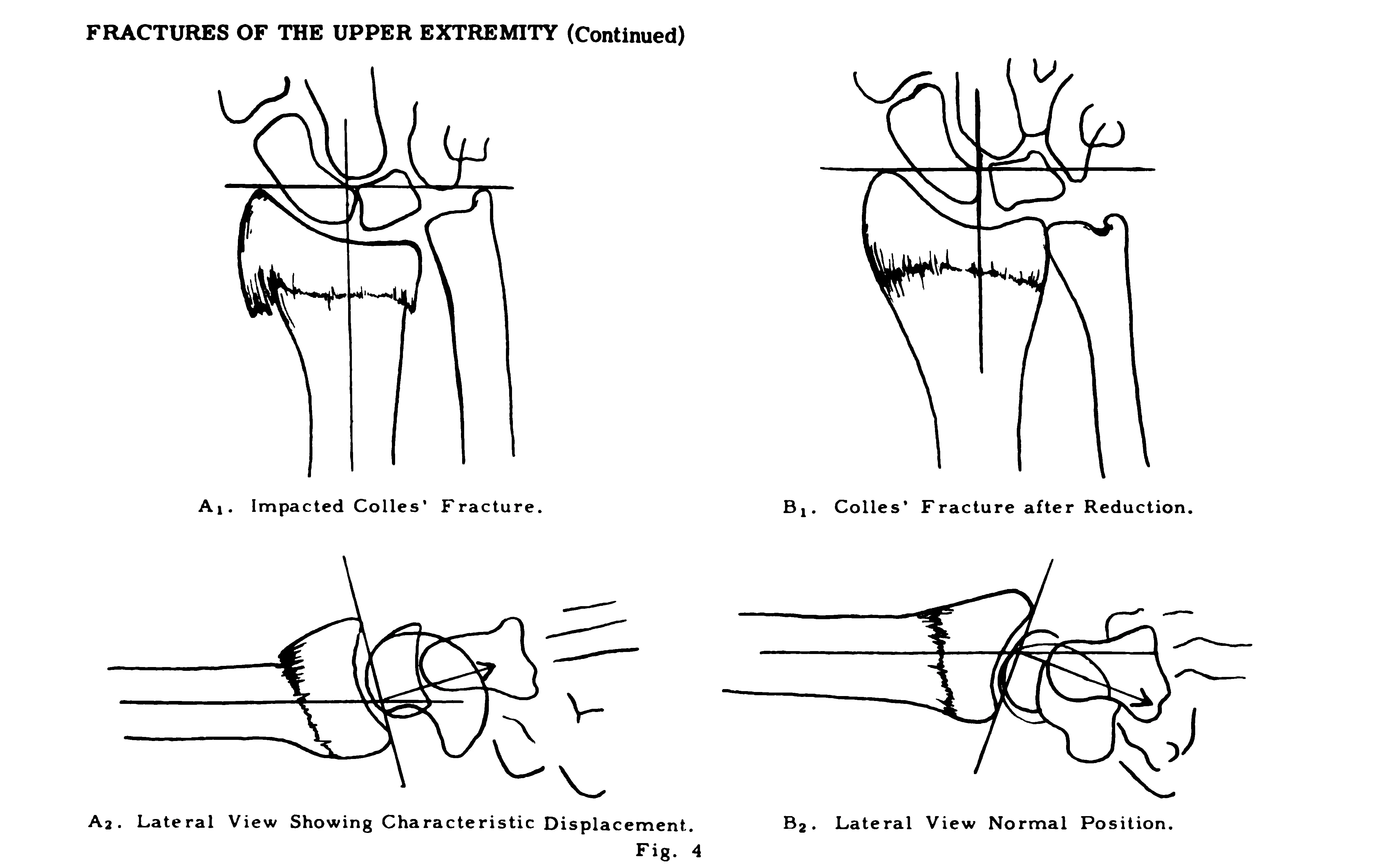
A fracture is a broken bone. With an open (or compound) fracture, the skin is broken above the fracture. With a closed (or simple) fracture, the skin is not broken, and no wound is visible. Fractures can also be classified by the type of damage to the bone:
- Incomplete: The fracture line does not go across the entire cross section of the bone.
- Complete: The fracture line goes across the entire cross section of the bone.
- Displaced: The bone fragments are not in alignment.
- Nondisplaced: The bone fragments remain in alignment.
- Comminuted: The bone breaks into three or more fragments.
- Compression: The fracture results in the bone (e.g., vertebrae) collapsing.
- Greenstick: The bone breaks on one side and is bent but still intact on the other side; commonly seen in children.
- Impacted: The bone fragments are driven into each other.
- Linear or longitudinal: The fracture line runs along the length of the bone.
- Oblique: The fracture runs at an oblique angle across the bone.
- Pathologic (or spontaneous): The fracture is due to weakening of the bone structure by a pathologic condition.
- Spiral: The fracture line resembles a spiral; caused by the bone being twisted.
- Transverse: The fracture runs perpendicular across the bone.
Fractures can occur from trauma, overuse, or diseases. A pathologic fracture results from an underlying disease, such as osteoporosis or cancer. Signs and symptoms of a fracture include pain, swelling, bruising, numbness or tingling, deformity, and difficulty using or inability to use the affected area. Bleeding at the site of the break will also occur with compound fractures.
Most commonly, the provider will order an x-ray, but a bone scan, computed tomography (CT) scan, or magnetic resonance imaging (MRI) may also be used as a diagnostic test for some types of fractures. Depending on the type of fracture, treatment may include a cast, splint, or brace to immobilize the area. Surgery will be done if the bones are not aligned. During an open reduction and internal fixation (ORIF) surgery, the surgeon reduces (repositions and aligns) the bones and uses internal fixators (special metal devices [rods, screws, plates]) to hold the bones in place during the healing process. Sometimes, external fixators and traction are used to hold the bones in place. Medications may also be prescribed depending on the severity of the fracture. The provider may order analgesics for the pain, anti-inflammatory drugs, and muscle relaxants. Fractures may take several weeks to months to heal. Crutches or other assistive devices may be used for lower extremity fractures. A sling is used for an upper extremity fracture. Physical therapy may be ordered once the fracture is healed to strengthen the extremity.
Herniated disk
When one of the soft disks between the vertebrae has a crack, some of the jelly-like substance can leak out. This is called a ruptured, slipped, or herniated disk. The leaking jelly-like substance can lead to inflammation and compression of the local nerve.
The disks can degenerate with age. Middle-aged and older men are more at risk for a herniated disk. Typically, herniation occurs with strenuous activities, but it can also occur with lifting heavy objects, obesity, repetitive movements involving the lower back, long periods of sitting or standing, inactivity, and smoking. Signs and symptoms of a lower back herniated disk include sciatica (pain and numbness from the hip down to the foot), back pain, and weakness. The pain usually starts slowly and gets worse with standing or sitting. A herniated disk in the neck may cause pain and numbness from the shoulder down to the fingers.
The provider will check the patient’s reflexes and muscle strength. The provider may order diagnostic tests (e.g., myelogram and electromyography [EMG]) and imaging procedures (e.g., spine MRI, spine CT, and spine x-ray). The following are common treatments:
- Medications (e.g., nonsteroidal anti-inflammatory drugs [NSAIDs], narcotics, and muscle relaxants) to help reduce the pain, inflammation, and muscle spasms
- Rest and physical therapy
- Surgery to remove the herniation if the symptoms remain unchanged
Usually, the condition improves in 4 to 6 weeks.
Lordosis, kyphosis, and scoliosis
The spine has normal curves. Abnormal curvatures of the spine deprive the body of important features related to spinal strength, balance, and posture. The three most common curvatures are as follows:
- **Lordosis **(swayback): An exaggerated forward curve in the lumbar region, which makes the buttock appear larger
- **Kyphosis **(hunchback): An exaggerated curve in the thoracic region
- Scoliosis: An abnormal side-to-side (S or C) curvature that could be in both the thoracic and lumbar regions of the back
Lordosis is caused by genetic and congenital conditions (e.g., muscular dystrophy, spondylolisthesis). Kyphosis can occur at any age, but it more commonly occurs in adults due to degenerative diseases of the spine, fractures, and injury. Kyphosis can also be caused by Paget disease, muscular dystrophy, scoliosis, and tumors. Many times, scoliosis occurs in children from age 10 to early teens, and the cause is unknown. Abnormalities in any of the spinal curves can cause pain and fatigue in the lower back and legs. Lordosis can also cause pain that may affect mobility. Mild kyphosis may cause few symptoms, but severe kyphosis can cause back pain, stiffness, and an abnormally curved spine. Scoliosis can cause uneven shoulders and waist. One shoulder blade may be more prominent, and one hip may be higher than the other.
The provider will perform a physical exam of the spine and a neurologic exam. A spinal x-ray, an MRI, and CT scan are also used as diagnostic tools. The treatment depends on the severity of the curve and the patient’s age and condition. Treatments include physical therapy, medications (analgesics, NSAIDs, steroids, and muscle relaxants), spinal fusion surgery, and a back brace; surgical placement of metal stabilizing rods may be needed for scoliosis. In most cases, with treatment, the prognosis is good.
Osteomalacia and rickets
Osteomalacia is the softening of bones due to the lack of vitamin D. Vitamin D helps the body absorb calcium. Calcium is needed for strong bones. Osteomalacia occurs in adults and children; in children, this disease is called rickets.
Osteomalacia and rickets are caused by a lack of vitamin D, which can be related to an absorption issue (e.g., after gastric bypass surgery) or a lack of dietary intake. Osteomalacia can also be related to kidney failure, liver disease, and certain medications (e.g., anticonvulsants). Signs and symptoms include bone fractures without related injuries, muscle weakness, and widespread bone pain, usually in the hips.
The provider will order blood tests to check the vitamin D level, along with creatinine, electrolytes (e.g., calcium, phosphate), alkaline phosphatase, and parathyroid hormone levels. Bone x-rays, a bone density test, and a bone biopsy may be done. Treatment involves oral supplements of vitamin D, calcium, and phosphorus. Sunlight for vitamin D production is also recommended. With early treatment, the prognosis for rickets in children is excellent. The prognosis for osteomalacia is good if the vitamin D deficiency is addressed.
Osteoporosis
Osteoporosis is a condition that causes bones to become brittle and weak. Healthy bone is constantly being broken down and remodeled. When a person has osteoporosis, bones are being broken down more quickly than new bone can be created. This leads to weak bones that are easily fractured, especially in the wrist, hip, and spine.
As people age, bone mass is lost faster than it can be produced, leading to osteoporosis. Increased risks for osteoporosis include menopause, family history, small body frame, white or Asian descent, hormonal imbalance, dietary deficiencies (low calcium intake), long-term corticosteroid use, celiac disease, kidney or liver disease, cancer, and some autoimmune conditions. A sedentary lifestyle, tobacco use, and excessive alcohol consumption are also risk factors for osteoporosis. Early osteoporosis is asymptomatic, but as the bones become weaker, back pain, height loss, stooped posture, and fractures can occur.
A bone density test is the only diagnostic tool for osteoporosis. This low-level x-ray determines the mineral content of the bone. The following treatments are used for osteoporosis:
- Weight-bearing exercises to maintain bone density
- Modifying risk factors to prevent falls
- Appropriate calcium and vitamin D intake from food or supplements
- Medications, such as bisphosphonates and hormone replacement therapy (estrogen)
Osteoporosis is a chronic condition that can be managed but does progress with age.
Healthy, strong bones
Peak bone mass is usually achieved around age 30. After that time, a little more bone is lost through remodeling than what is gained. This leads to a decrease in bone mass or density. For good bone health, it is important to do the following:
- Eat a diet rich in calcium. Good sources of calcium include dairy products, canned salmon with bones, soy products (e.g., tofu), broccoli and kale, and almonds.
- Take in adequate amounts of vitamin D. Vitamin D helps the body absorb dietary phosphorus and calcium. Good dietary sources of vitamin D include oily fish (e.g., trout, tuna, salmon), mushrooms, eggs, and fortified cereals and milk. Sunlight also helps in the body’s production of vitamin D.
- Limit tobacco and alcohol use. Tobacco use can lead to weak bones. Drinking more than one alcoholic drink per day for women and two for men can increase bone density loss.
- Do weight-bearing exercises. Weight-bearing exercises, such as jogging, climbing stairs, and walking, help build strong bones and slow bone loss.