



Hematology in the reference laboratory
The reference laboratory performs coagulation testing, blood smear preparation and microscopic examination, and platelet indices. As mentioned in a prior section, the reference laboratory may complete the automated CBC or perform a manual CBC if abnormalities are indicated. The following sections discuss tests done in the reference laboratory.
Partial thromboplastin time
A PTT test measures the time it takes for a blood clot to form. The PTT test is used to check the function of specific coagulation factors. If any factors are missing, it can take longer for the blood to clot. A PTT test is also used to monitor patients taking heparin, an anticoagulant. The reference range of the PTT is 25 to 35 seconds. If a patient is receiving anticoagulant therapy, the reference range is 1.5 to 2.5 times the normal reference range.
With the activated partial thromboplastin time (aPTT) test, an activator is added, which speeds up the clotting time. The reference range of the aPTT is 25 to 35 seconds.
Another common coagulation test is the prothrombin time (PT), often reported with an international normalized ratio (INR). PT/INR monitors patients taking warfarin, while the aPTT monitors patients taking heparin.
Preparing and the examination of blood smears
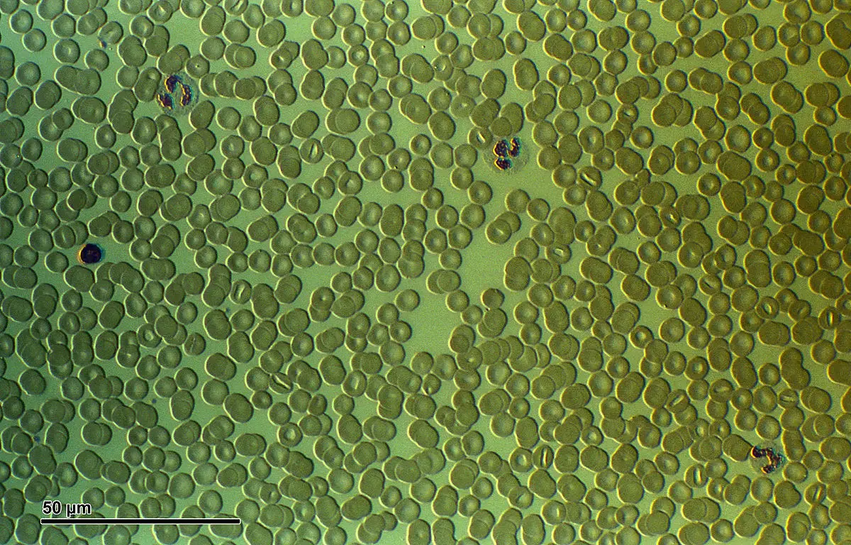
When blood must be microscopically examined, a blood smear needs to be created, and stain may be added. A blood smear enables the examiner to view the preserved cellular structures of blood. The morphology of WBCs, RBCs, and platelets can be studied. Their size, shape, and maturity can also be evaluated.
Preparing blood smears
A blood smear is prepared by placing a drop of blood from a fingerstick or an EDTA tube (using a DIFF-SAFE blood dispenser) onto a clean glass slide. The slide must be free of dust and grease. The best specimen for a blood smear is capillary blood that has no anticoagulant added. EDTA-anticoagulated blood can be used, provided the smear is made within 2 hours of specimen collection. Because timing is important, the medical assistant may be asked to prepare a smear during collection of the CBC specimen.
The wedge smear is used most frequently and is obtained by following these steps:
- A small drop of blood is placed ½ inch from the frosted end (placed to the right) of a glass slide.
- The end of a second glass spreader slide is placed in front (to the left) of the drop of blood at an angle of 30 to 35 degrees.
- The spreader slide is brought back into the drop with a quick but smooth gliding motion until the blood spreads along the edge of the spreader slide.
- The spreader slide is then pushed to the left with a quick, steady motion, spreading the blood across the slide.
A good wedge smear should cover one-half to three-quarters of the slide. It should show a gradual transition from a thick to a thin end with a feathered edge. It should have a smooth appearance with no ridges, holes, lines, streaks, or clumps. On microscopic examination, the cells should be evenly distributed.
After the smear has been made, the patient’s name is written on the frosted end of the slide with a pencil, and the smear is allowed to air-dry. The slide should be propped up to dry with the thick end down. Do not blow on the blood slide to dry it. The moisture in your breath can cause artifacts to form in the RBCs. Once dry, the slide is fixed, which preserves the cells and prevents changes in the cells. Many of the quick stains available on the market contain the fixative in the stain.
Staining blood smears
Stains commonly used to make blood smears are described as polychromatic. They contain dyes that stain cell components of different colors. These stains are attracted to different parts of the cell, which makes the cells and their structures easier to see and tell apart. The most commonly used differential blood stain is Wright’s stain.
Examination of a blood smear
It is important for the medical assistant to understand what occurs when blood smears are microscopically examined. Useful information can be gathered from a microscopic evaluation of blood cells in a stained smear.
Identification of normal blood cells
The three features hematologists look for in blood cells are the cell size, the appearance of the nucleus, and the appearance of the cytoplasm.
Differential examination
A specific area of a stained smear is examined under the microscope for a differential. The slide is examined near the feathered end of the smear, where cells are barely touching one another and are easiest to identify. Cells are examined with the oil immersion objective of the microscope. The differential involves counting and classifying 100 consecutive WBCs while moving in a winding pattern through the smear. A count of the cells observed is kept on a differential cell counter or a computer.
Red blood cell morphology
After the differential cell count has been determined, the RBCs are observed and evaluated. Normally, stained RBCs are the same size and shape and are well filled with hemoglobin. Any variations from the normal state are reported. The appearance of the RBCs should correlate with the RBC indices:
- Size: Normal-sized RBCs are said to be normocytic. If the cells are larger than normal, they are macrocytic. If the cells are smaller than normal, they are microcytic. The condition in which different sizes of RBCs are present is known as anisocytosis.
- Shape: Normal RBCs are round or slightly oval. Cells may be shaped like sickles, targets, crescents, or burs. Poikilocytosis is a significant variation in the shape of RBCs.
- Content: An RBC with a normal amount of hemoglobin is said to be normochromic. Pale-staining cells are hypochromic and have less hemoglobin than normal. Any inclusions within red blood cells should be reported.
Platelet analysis
On a stained smear, the platelets are observed for any abnormalities. Excessive platelet clumping is reported as part of the platelet analysis and may indicate a traumatic blood draw.
Platelet indices
With automated hematology analyzers, platelet indices may be reported as part of the CBC. Platelet indices are helpful to providers when they are diagnosing thromboembolic diseases. There are several platelet indices:
- Mean platelet volume (MPV): Measures the average size of the platelets in the blood sample. The normal range for an adult is 7.2 to 11.8 fL.
- Plateletcrit (Pct): Measures the total mass of platelets as a percentage volume in the blood. The normal range is 0.22% to 0.24%.
- Platelet distribution width (PDW): Measures the variation in the platelet size. The normal range for an adult is 10% to 18%.